

Transgenic Technology in Marine Organisms 13.3 Characterization of Transgenic Marine Organisms 397
Part B | 13.3
15
Southern blot analysis with transgene-specific probe
1 2 3 4 5 6 7 8 9 10 111213 14
16 17 18 19 20
PolyA + RNA pool
Primer 1
(transgene-specific)
Primer 2 (RACE-adaptor or
transgene-specific primer)
Oligo dT or 3'-RACE primer
PCR
Isolation of total RNA
Transgene transcript
1 st strand cDNA synthesis
1 st strand cDNA
PCR product
184 bp
b) Transgene expression in F 2 transgenic fish
a) Transgenic marine organisms
(A)n
(A)n
(A)n
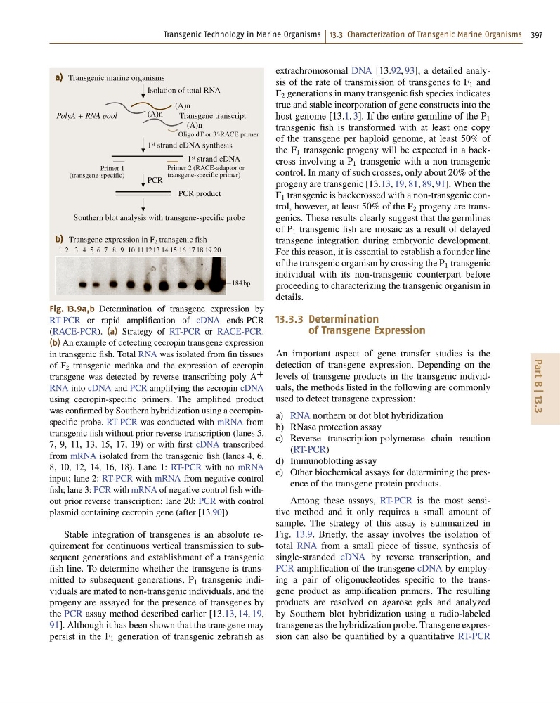
Fig. 13.9a,b Determination of transgene expression by
RT-PCR or rapid amplification of cDNA ends-PCR
(RACE-PCR). (a) Strategy of RT-PCR or RACE-PCR.
(b) An example of detecting cecropin transgene expression
in transgenic fish. Total RNA was isolated from fin tissues
of F 2 transgenic medaka and the expression of cecropin
transgene was detected by reverse transcribing poly A
C
RNA into cDNA and PCR amplifying the cecropin cDNA
using cecropin-specific primers. The amplified product
was confirmed by Southern hybridization using a cecropinspecific probe. RT-PCR was conducted with mRNA from
transgenic fish without prior reverse transcription (lanes 5,
7, 9, 11, 13, 15, 17, 19) or with first cDNA transcribed
from mRNA isolated from the transgenic fish (lanes 4, 6,
8, 10, 12, 14, 16, 18). Lane 1: RT-PCR with no mRNA
input; lane 2: RT-PCR with mRNA from negative control
fish; lane 3: PCR with mRNA of negative control fish without prior reverse transcription; lane 20: PCR with control
plasmid containing cecropin gene (after [13.90])
Stable integration of transgenes is an absolute requirement for continuous vertical transmission to subsequent generations and establishment of a transgenic
fish line. To determine whether the transgene is transmitted to subsequent generations, P 1 transgenic individuals are mated to non-transgenic individuals, and the
progeny are assayed for the presence of transgenes by
the PCR assay method described earlier [13.13, 14, 19,
91]. Although it has been shown that the transgene may
persist in the F 1 generation of transgenic zebrafish as
extrachromosomal DNA [13.92, 93], a detailed analysis of the rate of transmission of transgenes to F 1 and
F 2 generations in many transgenic fish species indicates
true and stable incorporation of gene constructs into the
host genome [13.1, 3]. If the entire germline of the P 1
transgenic fish is transformed with at least one copy
of the transgene per haploid genome, at least 50% of
the F 1 transgenic progeny will be expected in a backcross involving a P 1 transgenic with a non-transgenic
control. In many of such crosses, only about 20% of the
progeny are transgenic [13.13, 19, 81, 89, 91]. When the
F 1 transgenic is backcrossed with a non-transgenic control, however, at least 50% of the F 2 progeny are transgenics. These results clearly suggest that the germlines
of P 1 transgenic fish are mosaic as a result of delayed
transgene integration during embryonic development.
For this reason, it is essential to establish a founder line
of the transgenic organism by crossing the P 1 transgenic
individual with its non-transgenic counterpart before
proceeding to characterizing the transgenic organism in
details.
13.3.3 Determination
of Transgene Expression
An important aspect of gene transfer studies is the
detection of transgene expression. Depending on the
levels of transgene products in the transgenic individuals, the methods listed in the following are commonly
used to detect transgene expression:
a) RNA northern or dot blot hybridization
b) RNase protection assay
c) Reverse transcription-polymerase chain reaction
(RT-PCR)
d) Immunoblotting assay
e) Other biochemical assays for determining the presence of the transgene protein products.
Among these assays, RT-PCR is the most sensitive method and it only requires a small amount of
sample. The strategy of this assay is summarized in
Fig. 13.9. Briefly, the assay involves the isolation of
total RNA from a small piece of tissue, synthesis of
single-stranded cDNA by reverse transcription, and
PCR amplification of the transgene cDNA by employing a pair of oligonucleotides specific to the transgene product as amplification primers. The resulting
products are resolved on agarose gels and analyzed
by Southern blot hybridization using a radio-labeled
transgene as the hybridization probe. Transgene expression can also be quantified by a quantitative RT-PCR
Part B | 13.3
15
Southern blot analysis with transgene-specific probe
1 2 3 4 5 6 7 8 9 10 111213 14
16 17 18 19 20
PolyA + RNA pool
Primer 1
(transgene-specific)
Primer 2 (RACE-adaptor or
transgene-specific primer)
Oligo dT or 3'-RACE primer
PCR
Isolation of total RNA
Transgene transcript
1 st strand cDNA synthesis
1 st strand cDNA
PCR product
184 bp
b) Transgene expression in F 2 transgenic fish
a) Transgenic marine organisms
(A)n
(A)n
(A)n
Fig. 13.9a,b Determination of transgene expression by
RT-PCR or rapid amplification of cDNA ends-PCR
(RACE-PCR). (a) Strategy of RT-PCR or RACE-PCR.
(b) An example of detecting cecropin transgene expression
in transgenic fish. Total RNA was isolated from fin tissues
of F 2 transgenic medaka and the expression of cecropin
transgene was detected by reverse transcribing poly A
C
RNA into cDNA and PCR amplifying the cecropin cDNA
using cecropin-specific primers. The amplified product
was confirmed by Southern hybridization using a cecropinspecific probe. RT-PCR was conducted with mRNA from
transgenic fish without prior reverse transcription (lanes 5,
7, 9, 11, 13, 15, 17, 19) or with first cDNA transcribed
from mRNA isolated from the transgenic fish (lanes 4, 6,
8, 10, 12, 14, 16, 18). Lane 1: RT-PCR with no mRNA
input; lane 2: RT-PCR with mRNA from negative control
fish; lane 3: PCR with mRNA of negative control fish without prior reverse transcription; lane 20: PCR with control
plasmid containing cecropin gene (after [13.90])
Stable integration of transgenes is an absolute requirement for continuous vertical transmission to subsequent generations and establishment of a transgenic
fish line. To determine whether the transgene is transmitted to subsequent generations, P 1 transgenic individuals are mated to non-transgenic individuals, and the
progeny are assayed for the presence of transgenes by
the PCR assay method described earlier [13.13, 14, 19,
91]. Although it has been shown that the transgene may
persist in the F 1 generation of transgenic zebrafish as
extrachromosomal DNA [13.92, 93], a detailed analysis of the rate of transmission of transgenes to F 1 and
F 2 generations in many transgenic fish species indicates
true and stable incorporation of gene constructs into the
host genome [13.1, 3]. If the entire germline of the P 1
transgenic fish is transformed with at least one copy
of the transgene per haploid genome, at least 50% of
the F 1 transgenic progeny will be expected in a backcross involving a P 1 transgenic with a non-transgenic
control. In many of such crosses, only about 20% of the
progeny are transgenic [13.13, 19, 81, 89, 91]. When the
F 1 transgenic is backcrossed with a non-transgenic control, however, at least 50% of the F 2 progeny are transgenics. These results clearly suggest that the germlines
of P 1 transgenic fish are mosaic as a result of delayed
transgene integration during embryonic development.
For this reason, it is essential to establish a founder line
of the transgenic organism by crossing the P 1 transgenic
individual with its non-transgenic counterpart before
proceeding to characterizing the transgenic organism in
details.
13.3.3 Determination
of Transgene Expression
An important aspect of gene transfer studies is the
detection of transgene expression. Depending on the
levels of transgene products in the transgenic individuals, the methods listed in the following are commonly
used to detect transgene expression:
a) RNA northern or dot blot hybridization
b) RNase protection assay
c) Reverse transcription-polymerase chain reaction
(RT-PCR)
d) Immunoblotting assay
e) Other biochemical assays for determining the presence of the transgene protein products.
Among these assays, RT-PCR is the most sensitive method and it only requires a small amount of
sample. The strategy of this assay is summarized in
Fig. 13.9. Briefly, the assay involves the isolation of
total RNA from a small piece of tissue, synthesis of
single-stranded cDNA by reverse transcription, and
PCR amplification of the transgene cDNA by employing a pair of oligonucleotides specific to the transgene product as amplification primers. The resulting
products are resolved on agarose gels and analyzed
by Southern blot hybridization using a radio-labeled
transgene as the hybridization probe. Transgene expression can also be quantified by a quantitative RT-PCR