

Transgenic Technology in Marine Organisms 13.2 Production of Transgenic Marine Organisms 393
Part B | 13.2
Table 13.2 Parameters of fish transgenesis by microinjection and electroporation
Parameters
Gene transfer method
Microinjection
Electroporation
Developmental
stage
1–2 cells
1–2 cells
DNA size
< 10 Kb
< 10 Kb
DNA
concentration
10 67 molecules/
embryo 100 g=mL
DNA topology
linear
linear
Chorion barrier
dechorionated/
micropyle
intact chorion
Electrical field
strength
N/A a
5003000 V
Pulse shape
N/A a
exponential/
square
Pulse duration
N/A a
ms c to s
Temperature
RT b
RT b
Medium
PBS/saline
PBS/saline
a N/A, not applicable, b RT – room temperature (25 ı C), c ms –
millisecond. These parameters were extracted from the following references [13.5, 11, 13, 15]
justing the photo-period and the water temperature,
precisely staged newly fertilized eggs can be collected
from the aquaria for gene transfer. If medaka eggs are
maintained at 4
ı C immediately after fertilization, the
micropyle on the fertilized eggs will remain visible at
least for 2 h. The DNA solution can be delivered into
the embryos by injection through the micropyle during
this time period.
Depending on the fish species, the survival rate of
injected fish embryos ranges from 35 to 80%, while
the rate of DNA integration ranges from 10 to 70% in
the survivors [13.1, 2]. The tough chorions of the fertilized eggs in some fish species, e.g., rainbow trout and
Atlantic salmon, can make insertion of glass needles
difficult. This difficulty has been overcome by employing any one of the following methods:
i) Inserting the injection needles through the micropyle
ii) Making an opening on the egg chorions by microsurgery
iii) Removing the chorion by mechanical or enzymatic
means
iv) Preventing chorion hardening by initiating fertilization in a solution containing 1 mM glutathione
v) Injecting the unfertilized eggs directly.
Electroporation
Electroporation is a successful method for transferring
foreign DNA into bacteria, yeast, plant, and animal
cells in culture [13.67–69]. This method has become
popular for transferring foreign genes into fish embryos (or sperm) or newly fertilized eggs of dwarf
surf clam in the past few years [13.5, 11, 70, 71]. The
principle of electroporation is utilizing a series of
short electrical pulses to permeate cell membranes,
thereby permitting the entry of DNA molecules into
embryos or sperm. The patterns of electrical pulses
can be emitted in a single pulse of exponential decay
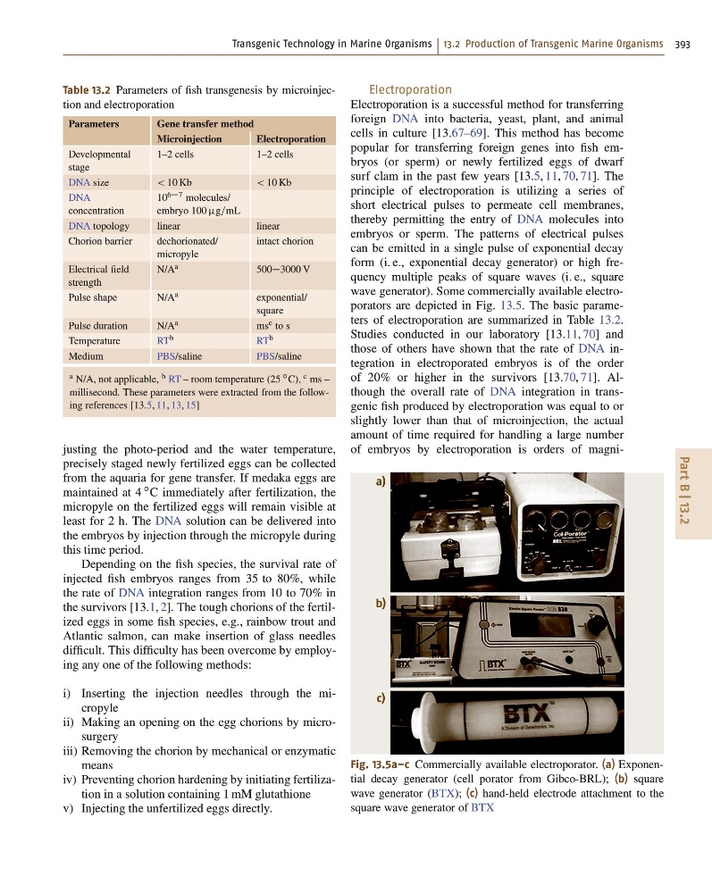
form (i. e., exponential decay generator) or high frequency multiple peaks of square waves (i. e., square
wave generator). Some commercially available electroporators are depicted in Fig. 13.5. The basic parameters of electroporation are summarized in Table 13.2.
Studies conducted in our laboratory [13.11, 70] and
those of others have shown that the rate of DNA integration in electroporated embryos is of the order
of 20% or higher in the survivors [13.70, 71]. Although the overall rate of DNA integration in transgenic fish produced by electroporation was equal to or
slightly lower than that of microinjection, the actual
amount of time required for handling a large number
of embryos by electroporation is orders of magnia)
b)
c)
Fig. 13.5a–c Commercially available electroporator. (a) Exponential decay generator (cell porator from Gibco-BRL); (b) square
wave generator (BTX); (c) hand-held electrode attachment to the
square wave generator of BTX
Part B | 13.2
Table 13.2 Parameters of fish transgenesis by microinjection and electroporation
Parameters
Gene transfer method
Microinjection
Electroporation
Developmental
stage
1–2 cells
1–2 cells
DNA size
< 10 Kb
< 10 Kb
DNA
concentration
10 67 molecules/
embryo 100 g=mL
DNA topology
linear
linear
Chorion barrier
dechorionated/
micropyle
intact chorion
Electrical field
strength
N/A a
5003000 V
Pulse shape
N/A a
exponential/
square
Pulse duration
N/A a
ms c to s
Temperature
RT b
RT b
Medium
PBS/saline
PBS/saline
a N/A, not applicable, b RT – room temperature (25 ı C), c ms –
millisecond. These parameters were extracted from the following references [13.5, 11, 13, 15]
justing the photo-period and the water temperature,
precisely staged newly fertilized eggs can be collected
from the aquaria for gene transfer. If medaka eggs are
maintained at 4
ı C immediately after fertilization, the
micropyle on the fertilized eggs will remain visible at
least for 2 h. The DNA solution can be delivered into
the embryos by injection through the micropyle during
this time period.
Depending on the fish species, the survival rate of
injected fish embryos ranges from 35 to 80%, while
the rate of DNA integration ranges from 10 to 70% in
the survivors [13.1, 2]. The tough chorions of the fertilized eggs in some fish species, e.g., rainbow trout and
Atlantic salmon, can make insertion of glass needles
difficult. This difficulty has been overcome by employing any one of the following methods:
i) Inserting the injection needles through the micropyle
ii) Making an opening on the egg chorions by microsurgery
iii) Removing the chorion by mechanical or enzymatic
means
iv) Preventing chorion hardening by initiating fertilization in a solution containing 1 mM glutathione
v) Injecting the unfertilized eggs directly.
Electroporation
Electroporation is a successful method for transferring
foreign DNA into bacteria, yeast, plant, and animal
cells in culture [13.67–69]. This method has become
popular for transferring foreign genes into fish embryos (or sperm) or newly fertilized eggs of dwarf
surf clam in the past few years [13.5, 11, 70, 71]. The
principle of electroporation is utilizing a series of
short electrical pulses to permeate cell membranes,
thereby permitting the entry of DNA molecules into
embryos or sperm. The patterns of electrical pulses
can be emitted in a single pulse of exponential decay
form (i. e., exponential decay generator) or high frequency multiple peaks of square waves (i. e., square
wave generator). Some commercially available electroporators are depicted in Fig. 13.5. The basic parameters of electroporation are summarized in Table 13.2.
Studies conducted in our laboratory [13.11, 70] and
those of others have shown that the rate of DNA integration in electroporated embryos is of the order
of 20% or higher in the survivors [13.70, 71]. Although the overall rate of DNA integration in transgenic fish produced by electroporation was equal to or
slightly lower than that of microinjection, the actual
amount of time required for handling a large number
of embryos by electroporation is orders of magnia)
b)
c)
Fig. 13.5a–c Commercially available electroporator. (a) Exponential decay generator (cell porator from Gibco-BRL); (b) square
wave generator (BTX); (c) hand-held electrode attachment to the
square wave generator of BTX