

Part B | 13.2
392 Part B Tools and Methods in Marine Biotechnology
Archaeospores
Thallus
Carpogonia Spermatia
Carpospores
Bladelet
Conchocelis
Conchospores
Conchosporangia
G a m e to p h y te ( 1 N )
S p o r o p h y te ( 2 N )
Fig. 13.3 Life cycle of Porphyra
about 1–2 years. Thus it will prolong inheritance studies. In addition, since these fish species have only
a single spawning cycle per year, this restricts the number of gene transfer attempt that can be conducted per
year.
Marine macroalgae, such as Porphyra and Laminaria, have a biphasic life cycle that alternates between
macroscopic, foliose, gametophytic phase, and microscopic, shell-boring, sporophytic filaments referred to
a)
b)
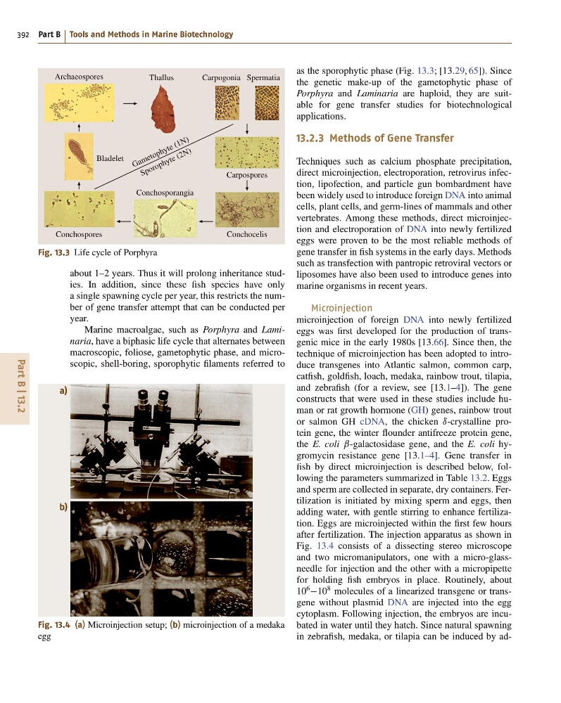
Fig. 13.4 (a) Microinjection setup; (b) microinjection of a medaka
egg
as the sporophytic phase (Fig. 13.3; [13.29, 65]). Since
the genetic make-up of the gametophytic phase of
Porphyra and Laminaria are haploid, they are suitable for gene transfer studies for biotechnological
applications.
13.2.3 Methods of Gene Transfer
Techniques such as calcium phosphate precipitation,
direct microinjection, electroporation, retrovirus infection, lipofection, and particle gun bombardment have
been widely used to introduce foreign DNA into animal
cells, plant cells, and germ-lines of mammals and other
vertebrates. Among these methods, direct microinjection and electroporation of DNA into newly fertilized
eggs were proven to be the most reliable methods of
gene transfer in fish systems in the early days. Methods
such as transfection with pantropic retroviral vectors or
liposomes have also been used to introduce genes into
marine organisms in recent years.
Microinjection
microinjection of foreign DNA into newly fertilized
eggs was first developed for the production of transgenic mice in the early 1980s [13.66]. Since then, the
technique of microinjection has been adopted to introduce transgenes into Atlantic salmon, common carp,
catfish, goldfish, loach, medaka, rainbow trout, tilapia,
and zebrafish (for a review, see [13.1–4]). The gene
constructs that were used in these studies include human or rat growth hormone (GH) genes, rainbow trout
or salmon GH cDNA, the chicken ı-crystalline protein gene, the winter flounder antifreeze protein gene,
the E. coli ˇ-galactosidase gene, and the E. coli hygromycin resistance gene [13.1–4]. Gene transfer in
fish by direct microinjection is described below, following the parameters summarized in Table 13.2. Eggs
and sperm are collected in separate, dry containers. Fertilization is initiated by mixing sperm and eggs, then
adding water, with gentle stirring to enhance fertilization. Eggs are microinjected within the first few hours
after fertilization. The injection apparatus as shown in
Fig. 13.4 consists of a dissecting stereo microscope
and two micromanipulators, one with a micro-glassneedle for injection and the other with a micropipette
for holding fish embryos in place. Routinely, about
10
6
10
8 molecules of a linearized transgene or transgene without plasmid DNA are injected into the egg
cytoplasm. Following injection, the embryos are incubated in water until they hatch. Since natural spawning
in zebrafish, medaka, or tilapia can be induced by ad-
392 Part B Tools and Methods in Marine Biotechnology
Archaeospores
Thallus
Carpogonia Spermatia
Carpospores
Bladelet
Conchocelis
Conchospores
Conchosporangia
G a m e to p h y te ( 1 N )
S p o r o p h y te ( 2 N )
Fig. 13.3 Life cycle of Porphyra
about 1–2 years. Thus it will prolong inheritance studies. In addition, since these fish species have only
a single spawning cycle per year, this restricts the number of gene transfer attempt that can be conducted per
year.
Marine macroalgae, such as Porphyra and Laminaria, have a biphasic life cycle that alternates between
macroscopic, foliose, gametophytic phase, and microscopic, shell-boring, sporophytic filaments referred to
a)
b)
Fig. 13.4 (a) Microinjection setup; (b) microinjection of a medaka
egg
as the sporophytic phase (Fig. 13.3; [13.29, 65]). Since
the genetic make-up of the gametophytic phase of
Porphyra and Laminaria are haploid, they are suitable for gene transfer studies for biotechnological
applications.
13.2.3 Methods of Gene Transfer
Techniques such as calcium phosphate precipitation,
direct microinjection, electroporation, retrovirus infection, lipofection, and particle gun bombardment have
been widely used to introduce foreign DNA into animal
cells, plant cells, and germ-lines of mammals and other
vertebrates. Among these methods, direct microinjection and electroporation of DNA into newly fertilized
eggs were proven to be the most reliable methods of
gene transfer in fish systems in the early days. Methods
such as transfection with pantropic retroviral vectors or
liposomes have also been used to introduce genes into
marine organisms in recent years.
Microinjection
microinjection of foreign DNA into newly fertilized
eggs was first developed for the production of transgenic mice in the early 1980s [13.66]. Since then, the
technique of microinjection has been adopted to introduce transgenes into Atlantic salmon, common carp,
catfish, goldfish, loach, medaka, rainbow trout, tilapia,
and zebrafish (for a review, see [13.1–4]). The gene
constructs that were used in these studies include human or rat growth hormone (GH) genes, rainbow trout
or salmon GH cDNA, the chicken ı-crystalline protein gene, the winter flounder antifreeze protein gene,
the E. coli ˇ-galactosidase gene, and the E. coli hygromycin resistance gene [13.1–4]. Gene transfer in
fish by direct microinjection is described below, following the parameters summarized in Table 13.2. Eggs
and sperm are collected in separate, dry containers. Fertilization is initiated by mixing sperm and eggs, then
adding water, with gentle stirring to enhance fertilization. Eggs are microinjected within the first few hours
after fertilization. The injection apparatus as shown in
Fig. 13.4 consists of a dissecting stereo microscope
and two micromanipulators, one with a micro-glassneedle for injection and the other with a micropipette
for holding fish embryos in place. Routinely, about
10
6
10
8 molecules of a linearized transgene or transgene without plasmid DNA are injected into the egg
cytoplasm. Following injection, the embryos are incubated in water until they hatch. Since natural spawning
in zebrafish, medaka, or tilapia can be induced by ad-