

Bioinformatic Techniques on Marine Genomics 10.4 Chemical Analysis of Volatile Microbial Metabolites 297
Part B | 10.4
10.2 Marine and Bacterial Fluorescence Shining Light
on Biological Questions
One of the most widespread and remarkable analytical
techniques to be used in biology in recent years is the
use of a green fluorescent protein (GFP) from a jellyfish
(Fig. 10.2) to tag and study biological molecules under
the microscope in vivo. The natural function of GFP
in the jellyfish is unknown, although it has been suggested that the protein may act as a light driven electron
transfer. Different GFP-like proteins have been discovered in a variety of marine organisms, including the
marine chordate Amphioxus, underlying marine biodiversity as a rich source of biologically useful molecules
which can have a major impact on the development
of new analytical methods. It will be interesting to
monitor the discovery of new fluorescent proteins from
bacteria which may provide better markers or markers that allow the visualization of different processes
(e.g., redox state) to be visualized in addition to the
localization of specific proteins. One example is the
development of fluorescent proteins as markers which
emit light in the infrared region of the electromagnetic spectrum, allowing visualization of processes that
occur deeper in tissues, which are impenetrable to visible light. One of these is an engineered phytochrome,
named IPF1.4 from the bacterium Deinococcus radiodurans, a bacterium which is highly resistant to
ionizing radiation. More recently, the photosynthetic
bacterium Rhodopseudomonas palustris has provided
a phytochrome, which has been engineered to produce
the protein near-infrared fluorescent protein (iRFP for
improved infrared imaging.
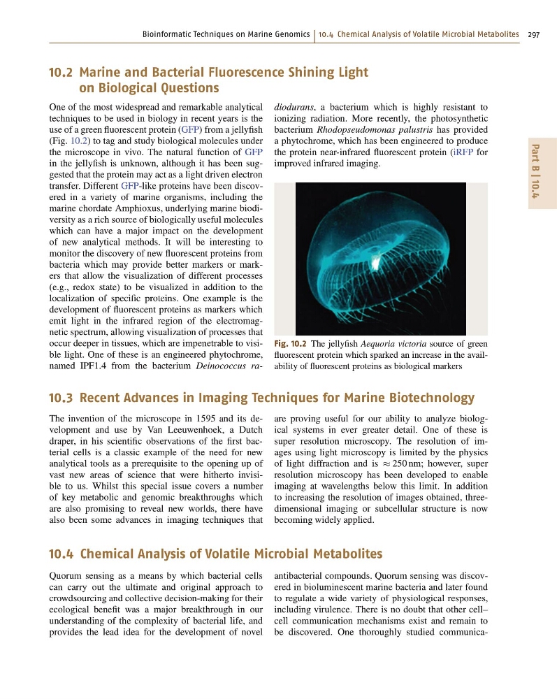
Fig. 10.2 The jellyfish Aequoria victoria source of green
fluorescent protein which sparked an increase in the availability of fluorescent proteins as biological markers
10.3 Recent Advances in Imaging Techniques for Marine Biotechnology
The invention of the microscope in 1595 and its development and use by Van Leeuwenhoek, a Dutch
draper, in his scientific observations of the first bacterial cells is a classic example of the need for new
analytical tools as a prerequisite to the opening up of
vast new areas of science that were hitherto invisible to us. Whilst this special issue covers a number
of key metabolic and genomic breakthroughs which
are also promising to reveal new worlds, there have
also been some advances in imaging techniques that
are proving useful for our ability to analyze biological systems in ever greater detail. One of these is
super resolution microscopy. The resolution of images using light microscopy is limited by the physics
of light diffraction and is 250 nm; however, super
resolution microscopy has been developed to enable
imaging at wavelengths below this limit. In addition
to increasing the resolution of images obtained, threedimensional imaging or subcellular structure is now
becoming widely applied.
10.4 Chemical Analysis of Volatile Microbial Metabolites
Quorum sensing as a means by which bacterial cells
can carry out the ultimate and original approach to
crowdsourcing and collective decision-making for their
ecological benefit was a major breakthrough in our
understanding of the complexity of bacterial life, and
provides the lead idea for the development of novel
antibacterial compounds. Quorum sensing was discovered in bioluminescent marine bacteria and later found
to regulate a wide variety of physiological responses,
including virulence. There is no doubt that other cell–
cell communication mechanisms exist and remain to
be discovered. One thoroughly studied communica-
Part B | 10.4
10.2 Marine and Bacterial Fluorescence Shining Light
on Biological Questions
One of the most widespread and remarkable analytical
techniques to be used in biology in recent years is the
use of a green fluorescent protein (GFP) from a jellyfish
(Fig. 10.2) to tag and study biological molecules under
the microscope in vivo. The natural function of GFP
in the jellyfish is unknown, although it has been suggested that the protein may act as a light driven electron
transfer. Different GFP-like proteins have been discovered in a variety of marine organisms, including the
marine chordate Amphioxus, underlying marine biodiversity as a rich source of biologically useful molecules
which can have a major impact on the development
of new analytical methods. It will be interesting to
monitor the discovery of new fluorescent proteins from
bacteria which may provide better markers or markers that allow the visualization of different processes
(e.g., redox state) to be visualized in addition to the
localization of specific proteins. One example is the
development of fluorescent proteins as markers which
emit light in the infrared region of the electromagnetic spectrum, allowing visualization of processes that
occur deeper in tissues, which are impenetrable to visible light. One of these is an engineered phytochrome,
named IPF1.4 from the bacterium Deinococcus radiodurans, a bacterium which is highly resistant to
ionizing radiation. More recently, the photosynthetic
bacterium Rhodopseudomonas palustris has provided
a phytochrome, which has been engineered to produce
the protein near-infrared fluorescent protein (iRFP for
improved infrared imaging.
Fig. 10.2 The jellyfish Aequoria victoria source of green
fluorescent protein which sparked an increase in the availability of fluorescent proteins as biological markers
10.3 Recent Advances in Imaging Techniques for Marine Biotechnology
The invention of the microscope in 1595 and its development and use by Van Leeuwenhoek, a Dutch
draper, in his scientific observations of the first bacterial cells is a classic example of the need for new
analytical tools as a prerequisite to the opening up of
vast new areas of science that were hitherto invisible to us. Whilst this special issue covers a number
of key metabolic and genomic breakthroughs which
are also promising to reveal new worlds, there have
also been some advances in imaging techniques that
are proving useful for our ability to analyze biological systems in ever greater detail. One of these is
super resolution microscopy. The resolution of images using light microscopy is limited by the physics
of light diffraction and is 250 nm; however, super
resolution microscopy has been developed to enable
imaging at wavelengths below this limit. In addition
to increasing the resolution of images obtained, threedimensional imaging or subcellular structure is now
becoming widely applied.
10.4 Chemical Analysis of Volatile Microbial Metabolites
Quorum sensing as a means by which bacterial cells
can carry out the ultimate and original approach to
crowdsourcing and collective decision-making for their
ecological benefit was a major breakthrough in our
understanding of the complexity of bacterial life, and
provides the lead idea for the development of novel
antibacterial compounds. Quorum sensing was discovered in bioluminescent marine bacteria and later found
to regulate a wide variety of physiological responses,
including virulence. There is no doubt that other cell–
cell communication mechanisms exist and remain to
be discovered. One thoroughly studied communica-