

224
I.R. Cowan
(a)
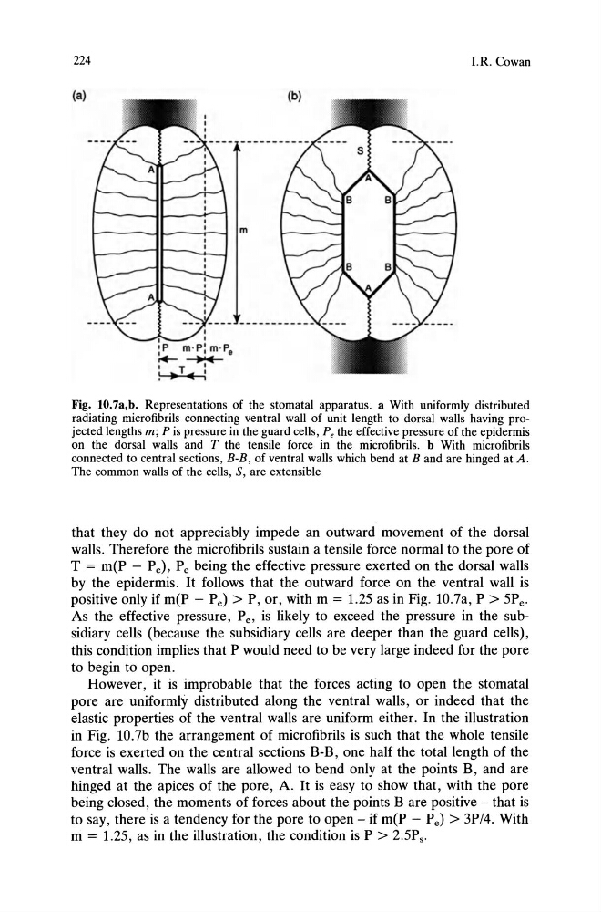
Fig. lO.7a,b. Representations of the stomatal apparatus. a With uniformly distributed
radiating microfibrils connecting ventral wall of unit length to dorsal walls having projected lengths m; P is pressure in the guard cells, Pe the effective pressure of the epidermis
on the dorsal walls and T the tensile force in the microfibrils. b With microfibrils
connected to central sections, B-B, of ventral walls which bend at B and are hinged at A.
The common walls of the cells, S, are extensible
that they do not appreciably impede an outward movement of the dorsal
walls. Therefore the micro fibrils sustain a tensile force normal to the pore of
T = m(P - P e), P e being the effective pressure exerted on the dorsal walls
by the epidermis. It follows that the outward force on the ventral wall is
positive only if m(P - Pe) > P, or, with m = 1.25 as in Fig. 1O.7a, P > 5Pe .
As the effective pressure, Pe , is likely to exceed the pressure in the subsidiary cells (because the subsidiary cells are deeper than the guard cells),
this condition implies that P would need to be very large indeed for the pore
to begin to open.
However, it is improbable that the forces acting to open the stomatal
pore are uniformly distributed along the ventral walls, or indeed that the
elastic properties of the ventral walls are uniform either. In the illustration
in Fig. lO.7b the arrangement of microfibrils is such that the whole tensile
force is exerted on the central sections B-B, one half the total length of the
ventral walls. The walls are allowed to bend only at the points B, and are
hinged at the apices of the pore, A. It is easy to show that, with the pore
being closed, the moments of forces about the points B are positive - that is
to say, there is a tendency for the pore to open - if m(P - Pe) > 3P/4. With
m = 1.25, as in the illustration, the condition is P > 2.5Ps .
I.R. Cowan
(a)
Fig. lO.7a,b. Representations of the stomatal apparatus. a With uniformly distributed
radiating microfibrils connecting ventral wall of unit length to dorsal walls having projected lengths m; P is pressure in the guard cells, Pe the effective pressure of the epidermis
on the dorsal walls and T the tensile force in the microfibrils. b With microfibrils
connected to central sections, B-B, of ventral walls which bend at B and are hinged at A.
The common walls of the cells, S, are extensible
that they do not appreciably impede an outward movement of the dorsal
walls. Therefore the micro fibrils sustain a tensile force normal to the pore of
T = m(P - P e), P e being the effective pressure exerted on the dorsal walls
by the epidermis. It follows that the outward force on the ventral wall is
positive only if m(P - Pe) > P, or, with m = 1.25 as in Fig. 1O.7a, P > 5Pe .
As the effective pressure, Pe , is likely to exceed the pressure in the subsidiary cells (because the subsidiary cells are deeper than the guard cells),
this condition implies that P would need to be very large indeed for the pore
to begin to open.
However, it is improbable that the forces acting to open the stomatal
pore are uniformly distributed along the ventral walls, or indeed that the
elastic properties of the ventral walls are uniform either. In the illustration
in Fig. lO.7b the arrangement of microfibrils is such that the whole tensile
force is exerted on the central sections B-B, one half the total length of the
ventral walls. The walls are allowed to bend only at the points B, and are
hinged at the apices of the pore, A. It is easy to show that, with the pore
being closed, the moments of forces about the points B are positive - that is
to say, there is a tendency for the pore to open - if m(P - Pe) > 3P/4. With
m = 1.25, as in the illustration, the condition is P > 2.5Ps .