

As to the Mode of Action of the Guard Cells in Dry Air
223
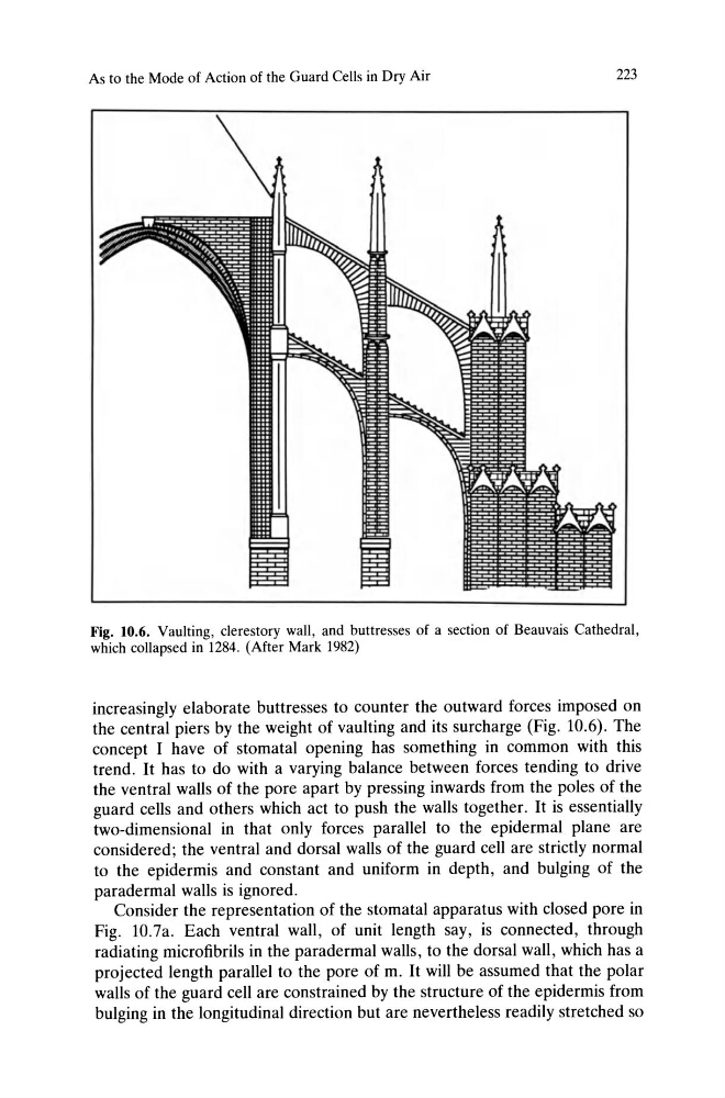
Fig. 10.6. Vaulting, clerestory wall, and buttresses of a section of Beauvais Cathedral,
which collapsed in 1284. (After Mark 1982)
increasingly elaborate buttresses to counter the outward forces imposed on
the central piers by the weight of vaulting and its surcharge (Fig. 10.6). The
concept I have of stomatal opening has something in common with this
trend. It has to do with a varying balance between forces tending to drive
the ventral walls of the pore apart by pressing inwards from the poles of the
guard cells and others which act to push the walls together. It is essentially
two-dimensional in that only forces parallel to the epidermal plane are
considered; the ventral and dorsal walls of the guard cell are strictly normal
to the epidermis and constant and uniform in depth, and bulging of the
paradermal walls is ignored.
Consider the representation of the stomatal apparatus with closed pore in
Fig. 1O.7a. Each ventral wall, of unit length say, is connected, through
radiating microfibrils in the paradermal walls, to the dorsal wall, which has a
projected length parallel to the pore of m. It will be assumed that the polar
walls of the guard cell are constrained by the structure of the epidermis from
bulging in the longitudinal direction but are nevertheless readily stretched so
223
Fig. 10.6. Vaulting, clerestory wall, and buttresses of a section of Beauvais Cathedral,
which collapsed in 1284. (After Mark 1982)
increasingly elaborate buttresses to counter the outward forces imposed on
the central piers by the weight of vaulting and its surcharge (Fig. 10.6). The
concept I have of stomatal opening has something in common with this
trend. It has to do with a varying balance between forces tending to drive
the ventral walls of the pore apart by pressing inwards from the poles of the
guard cells and others which act to push the walls together. It is essentially
two-dimensional in that only forces parallel to the epidermal plane are
considered; the ventral and dorsal walls of the guard cell are strictly normal
to the epidermis and constant and uniform in depth, and bulging of the
paradermal walls is ignored.
Consider the representation of the stomatal apparatus with closed pore in
Fig. 1O.7a. Each ventral wall, of unit length say, is connected, through
radiating microfibrils in the paradermal walls, to the dorsal wall, which has a
projected length parallel to the pore of m. It will be assumed that the polar
walls of the guard cell are constrained by the structure of the epidermis from
bulging in the longitudinal direction but are nevertheless readily stretched so