

!liII Methods Useful in Genetics and Oncology
0.5
~ .. -
,
,
,
~
I
,
"0
,
- ..-..
,
"""
,
LL
0
I
'-"
"0
,
I
I
- " - Wild Type
I
I
-
Heterozygous
,
- - - Homozygous Mutant
-0.5
........ No DNA
40
50
60
70
Temperature (O C)
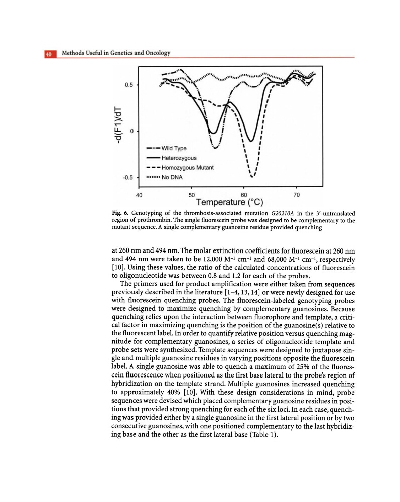
Fig. 6. Genotyping of the thrombosis-associated mutation G20210A in the 3'-untranslated
region of prothrombin. The single fluorescein probe was designed to be complementary to the
mutant sequence. A single complementary guanosine residue provided quenching
at 260 nm and 494 nm. The molar extinction coefficients for fluorescein at 260 nm
and 494 nm were taken to be 12,000 M-i cm- i and 68,000 M-i cm- i , respectively
[10]. Using these values, the ratio of the calculated concentrations of fluorescein
to oligonucleotide was between 0.8 and 1.2 for each of the probes.
The primers used for product amplification were either taken from sequences
previously described in the literature [1-4,13,14] or were newly designed for use
with fluorescein quenching probes. The fluorescein-labeled genotyping probes
were designed to maximize quenching by complementary guanosines. Because
quenching relies upon the interaction between fluorophore and template, a critical factor in maximizing quenching is the position of the guanosine(s) relative to
the fluorescent label. In order to quantify relative position versus quenching magnitude for complementary guanosines, a series of oligonucleotide template and
probe sets were synthesized. Template sequences were designed to juxtapose single and multiple guanosine residues in varying positions opposite the fluorescein
label. A single guanosine was able to quench a maximum of 25% of the fluorescein fluorescence when positioned as the first base lateral to the probe's region of
hybridization on the template strand. Multiple guanosines increased quenching
to approximately 40% [10]. With these design considerations in mind, probe
sequences were devised which placed complementary guanosine residues in positions that provided strong quenching for each of the six loci. In each case, quenching was provided either by a single guanosine in the first lateral position or by two
consecutive guanosines, with one positioned complementary to the last hybridizing base and the other as the first lateral base (Table 1).
0.5
~ .. -
,
,
,
~
I
,
"0
,
- ..-..
,
"""
,
LL
0
I
'-"
"0
,
I
I
- " - Wild Type
I
I
-
Heterozygous
,
- - - Homozygous Mutant
-0.5
........ No DNA
40
50
60
70
Temperature (O C)
Fig. 6. Genotyping of the thrombosis-associated mutation G20210A in the 3'-untranslated
region of prothrombin. The single fluorescein probe was designed to be complementary to the
mutant sequence. A single complementary guanosine residue provided quenching
at 260 nm and 494 nm. The molar extinction coefficients for fluorescein at 260 nm
and 494 nm were taken to be 12,000 M-i cm- i and 68,000 M-i cm- i , respectively
[10]. Using these values, the ratio of the calculated concentrations of fluorescein
to oligonucleotide was between 0.8 and 1.2 for each of the probes.
The primers used for product amplification were either taken from sequences
previously described in the literature [1-4,13,14] or were newly designed for use
with fluorescein quenching probes. The fluorescein-labeled genotyping probes
were designed to maximize quenching by complementary guanosines. Because
quenching relies upon the interaction between fluorophore and template, a critical factor in maximizing quenching is the position of the guanosine(s) relative to
the fluorescent label. In order to quantify relative position versus quenching magnitude for complementary guanosines, a series of oligonucleotide template and
probe sets were synthesized. Template sequences were designed to juxtapose single and multiple guanosine residues in varying positions opposite the fluorescein
label. A single guanosine was able to quench a maximum of 25% of the fluorescein fluorescence when positioned as the first base lateral to the probe's region of
hybridization on the template strand. Multiple guanosines increased quenching
to approximately 40% [10]. With these design considerations in mind, probe
sequences were devised which placed complementary guanosine residues in positions that provided strong quenching for each of the six loci. In each case, quenching was provided either by a single guanosine in the first lateral position or by two
consecutive guanosines, with one positioned complementary to the last hybridizing base and the other as the first lateral base (Table 1).