

Quantitative Two-Step RT-PCR for the Detection of Human ABCAI Transporter on LightCycier n
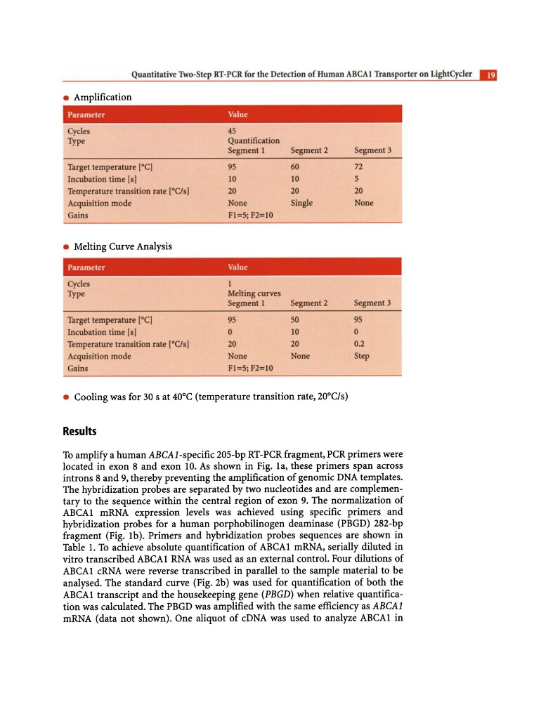
• Amplification
Parameter
Value
Cycles
Type
Target temperature [0C]
Incubation lime lsI
Temperature transition rate rOCls I
Acquisition mode
Gains
• Melting Curve Analysis
45
Quantification
Segment 1
95
10
20
None
FI=5; F2=10
Parameter
Value
Cycles
Type
Melting curves
Segment 1
Target temperature (OCI
95
Incubation time (sl
0
Temperature transition rate (OC/s]
20
Acquisition mode
None
Gains
FI=5;F2=10
Segment 2
60
10
20
Single
Segment 2
50
10
20
None
• Cooling was for 30 s at 40°C (temperature transition rate, 20°C/s)
Results
Segment 3
72
5
20
None
Segment 3
95
0
0.2
Step
To amplify a human ABCA l-specific 20S-bp RT -PCR fragment, PCR primers were
located in exon 8 and exon 10. As shown in Fig. la, these primers span across
introns 8 and 9, thereby preventing the amplification of genomic DNA templates.
The hybridization probes are separated by two nucleotides and are complementary to the sequence within the central region of exon 9. The normalization of
ABCA1 mRNA expression levels was achieved using specific primers and
hybridization probes for a human porphobilinogen deaminase (PBGD) 282-bp
fragment (Fig. 1b). Primers and hybridization probes sequences are shown in
Table 1. To achieve absolute quantification of ABCA1 mRNA, serially diluted in
vitro transcribed ABCA1 RNA was used as an external control. Four dilutions of
ABCA1 cRNA were reverse transcribed in parallel to the sample material to be
analysed. The standard curve (Fig. 2b) was used for quantification of both the
ABCA1 transcript and the housekeeping gene (PBGD) when relative quantification was calculated. The PBGD was amplified with the same efficiency as ABCAl
mRNA (data not shown). One aliquot of cDNA was used to analyze ABCA1 in
• Amplification
Parameter
Value
Cycles
Type
Target temperature [0C]
Incubation lime lsI
Temperature transition rate rOCls I
Acquisition mode
Gains
• Melting Curve Analysis
45
Quantification
Segment 1
95
10
20
None
FI=5; F2=10
Parameter
Value
Cycles
Type
Melting curves
Segment 1
Target temperature (OCI
95
Incubation time (sl
0
Temperature transition rate (OC/s]
20
Acquisition mode
None
Gains
FI=5;F2=10
Segment 2
60
10
20
Single
Segment 2
50
10
20
None
• Cooling was for 30 s at 40°C (temperature transition rate, 20°C/s)
Results
Segment 3
72
5
20
None
Segment 3
95
0
0.2
Step
To amplify a human ABCA l-specific 20S-bp RT -PCR fragment, PCR primers were
located in exon 8 and exon 10. As shown in Fig. la, these primers span across
introns 8 and 9, thereby preventing the amplification of genomic DNA templates.
The hybridization probes are separated by two nucleotides and are complementary to the sequence within the central region of exon 9. The normalization of
ABCA1 mRNA expression levels was achieved using specific primers and
hybridization probes for a human porphobilinogen deaminase (PBGD) 282-bp
fragment (Fig. 1b). Primers and hybridization probes sequences are shown in
Table 1. To achieve absolute quantification of ABCA1 mRNA, serially diluted in
vitro transcribed ABCA1 RNA was used as an external control. Four dilutions of
ABCA1 cRNA were reverse transcribed in parallel to the sample material to be
analysed. The standard curve (Fig. 2b) was used for quantification of both the
ABCA1 transcript and the housekeeping gene (PBGD) when relative quantification was calculated. The PBGD was amplified with the same efficiency as ABCAl
mRNA (data not shown). One aliquot of cDNA was used to analyze ABCA1 in