

Quantitative Analysis of AMLl-ETO Fusion Transcripts in t(8;21) Positive AML Using Real-Time RT-PCR 11m
10000000
1000000
100000
1 ()()()()
1 ()()()
100
10
,
\
A
~
\
\
\
\
\
/
/ '
Cytogenetic relapse
I- - AML1
/
/
I
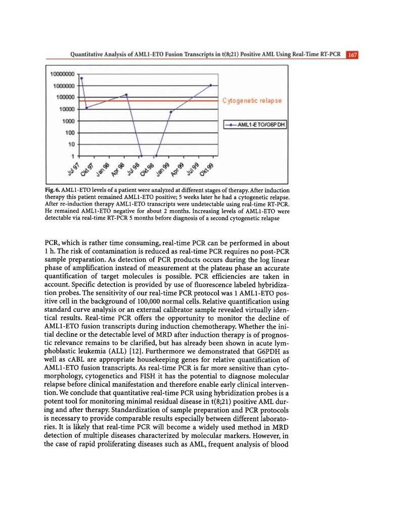
Fig. 6. AMLl-ETO levels of a patient were analyzed at different stages of therapy. After induction
therapy this patient remained AMLl-ETO positive; 5 weeks later he had a cytogenetic relapse.
After re-induction therapy AMLl-ETO transcripts were undetectable using real-time RT-PCR.
He remained AMLl-ETO negative for about 2 months. Increasing levels of AMLl-ETO were
detectable via real-time RT-PCR 5 months before diagnosis of a second cytogenetic relapse
peR, which is rather time consuming, real-time peR can be performed in about
1 h. The risk of contamination is reduced as real-time peR requires no post-peR
sample preparation. As detection of peR products occurs during the log linear
phase of amplification instead of measurement at the plateau phase an accurate
quantification of target molecules is possible. peR efficiencies are taken in
account. Specific detection is provided by use of fluorescence labeled hybridization probes. The sensitivity of our real-time peR protocol was 1 AMLl-ETO positive cell in the background of 100,000 normal cells. Relative quantification using
standard curve analysis or an external calibrator sample revealed virtually identical results. Real-time peR offers the opportunity to monitor the decline of
AMLl-ETO fusion transcripts during induction chemotherapy. Whether the initial decline or the detectable level of MRD after induction therapy is of prognostic relevance remains to be clarified, but has already been shown in acute lymphoblastic leukemia (ALL) [12]. Furthermore we demonstrated that G6PDH as
well as cABL are appropriate housekeeping genes for relative quantification of
AMLl-ETO fusion transcripts. As real-time peR is far more sensitive than cytomorphology, cytogenetics and FISH it has the potential to diagnose molecular
relapse before clinical manifestation and therefore enable early clinical intervention. We conclude that quantitative real-time peR using hybridization probes is a
potent tool for monitoring minimal residual disease in t(8;21) positive AML during and after therapy. Standardization of sample preparation and peR protocols
is necessary to provide comparable results especially between different laboratories. It is likely that real-time peR will become a widely used method in MRD
detection of multiple diseases characterized by molecular markers. However, in
the case of rapid proliferating diseases such as AML, frequent analysis of blood
10000000
1000000
100000
1 ()()()()
1 ()()()
100
10
,
\
A
~
\
\
\
\
\
/
/ '
Cytogenetic relapse
I- - AML1
/
/
I
Fig. 6. AMLl-ETO levels of a patient were analyzed at different stages of therapy. After induction
therapy this patient remained AMLl-ETO positive; 5 weeks later he had a cytogenetic relapse.
After re-induction therapy AMLl-ETO transcripts were undetectable using real-time RT-PCR.
He remained AMLl-ETO negative for about 2 months. Increasing levels of AMLl-ETO were
detectable via real-time RT-PCR 5 months before diagnosis of a second cytogenetic relapse
peR, which is rather time consuming, real-time peR can be performed in about
1 h. The risk of contamination is reduced as real-time peR requires no post-peR
sample preparation. As detection of peR products occurs during the log linear
phase of amplification instead of measurement at the plateau phase an accurate
quantification of target molecules is possible. peR efficiencies are taken in
account. Specific detection is provided by use of fluorescence labeled hybridization probes. The sensitivity of our real-time peR protocol was 1 AMLl-ETO positive cell in the background of 100,000 normal cells. Relative quantification using
standard curve analysis or an external calibrator sample revealed virtually identical results. Real-time peR offers the opportunity to monitor the decline of
AMLl-ETO fusion transcripts during induction chemotherapy. Whether the initial decline or the detectable level of MRD after induction therapy is of prognostic relevance remains to be clarified, but has already been shown in acute lymphoblastic leukemia (ALL) [12]. Furthermore we demonstrated that G6PDH as
well as cABL are appropriate housekeeping genes for relative quantification of
AMLl-ETO fusion transcripts. As real-time peR is far more sensitive than cytomorphology, cytogenetics and FISH it has the potential to diagnose molecular
relapse before clinical manifestation and therefore enable early clinical intervention. We conclude that quantitative real-time peR using hybridization probes is a
potent tool for monitoring minimal residual disease in t(8;21) positive AML during and after therapy. Standardization of sample preparation and peR protocols
is necessary to provide comparable results especially between different laboratories. It is likely that real-time peR will become a widely used method in MRD
detection of multiple diseases characterized by molecular markers. However, in
the case of rapid proliferating diseases such as AML, frequent analysis of blood