

Quantitative Analysis of AMLI-ETO Fusion Transcripts in t(8;2I) Positive AML Using Real-Time RT-PCR 1m
Procedure
Human blood and bone marrow samples were analyzed. Patients were selected
due to t(8;21) positive cytogenetics and were referred to our lab for central AML
diagnosis. Mononuclear cells were obtained by Ficoll Hypaque density gradient
centrifugation. Total RNA of 1 x 10 7 human cells and Kasumi 1 cells was isolated
according to the Qiagen RNeasy protocol. cDNA Synthesis was performed via
Boehringer Mannheim 1st Strand cDNA Synthesis Kit for RT-PCR (AMV) using
random hexamers, according to the manufacturer's protocol. cDNA of 10 6 cells
was then amplified using the LightCycler Faststart DNA Master. The PCR products were detected via specific fluorescence labeled hybridization probes. The
detection format is based on Fluorescence Resonance Energy Transfer (FRET)
[9,10]. To measure the sensitivity of our method we performed limiting dilution
experiments of Kasumi 1 cells in the background of mononuclear cells of a
healthy donor.
LightCycler data was analyzed using LightCycler 3.0 software and the second
derivative maximum method. Quantitative analysis was performed relative to
the housekeeping genes G6PDH and cABL [11, 12]. Therefore the ratio of target
gene to housekeeping gene was calculated. Relative quantification of AMLl-ETO
fusion transcripts was calculated according to standard curve analysis and in
parallel using a known concentration of an external calibrator. We used cDNA of
10 5 Kasumi 1 cells as an external calibrator and analyzed the transcript levels of
the unknown samples by using a ~-version of the relative quantification software. The AMLl-ETO level of the calibrator sample (l05 Kasumi 1 cells) was set
to a relative value of 1,000,000. PCR efficiencies for target and housekeeping
genes were measured by repeated generation of standard curves using dilution
series of Kasumi 1 cells and these efficiencies were used to calculate unknown
sample concentrations.
PCR was performed using 2 III mastermix (LightCycler Faststart DNA Master
Hybridization Probes, Roche Diagnostics, Mannheim, Germany), containing
buffer, dNTPs and Taq polymerase; 4 mM MgCL2; 0.25 IlM of each 3' and 5' fluorescent labeled hybridization probes (TIB Molbiol, Berlin, Germany); 0.5 IlM of
each 3'and 5' Primer (TIB Molbiol, Berlin, Germany), 2 III of cDNA and H 2 0 to a
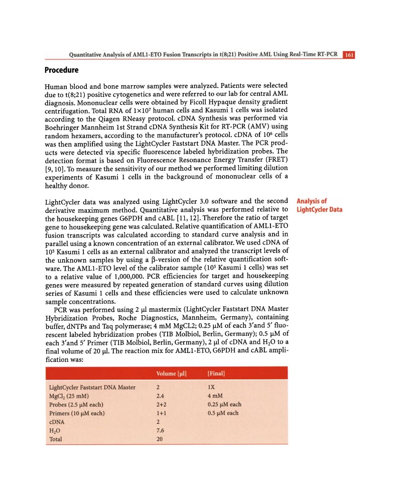
final volume of 20 jJI. The reaction mix for AMLl-ETO, G6PDH and cABL amplification was:
LightCyc1er Faststarl DNA Master
MgCI2 (25 mM)
Probes (2.5 f.LM each)
Primers (J 0 f.LM each)
eDNA
H20
Total
Volume [Jil l
[Finall
2
2.4
2+2
1+1
2
7.6
20
lX
4mM
0.25 f.LM each
0.5 f.LM each
Analysis of
lightCycier Data
Procedure
Human blood and bone marrow samples were analyzed. Patients were selected
due to t(8;21) positive cytogenetics and were referred to our lab for central AML
diagnosis. Mononuclear cells were obtained by Ficoll Hypaque density gradient
centrifugation. Total RNA of 1 x 10 7 human cells and Kasumi 1 cells was isolated
according to the Qiagen RNeasy protocol. cDNA Synthesis was performed via
Boehringer Mannheim 1st Strand cDNA Synthesis Kit for RT-PCR (AMV) using
random hexamers, according to the manufacturer's protocol. cDNA of 10 6 cells
was then amplified using the LightCycler Faststart DNA Master. The PCR products were detected via specific fluorescence labeled hybridization probes. The
detection format is based on Fluorescence Resonance Energy Transfer (FRET)
[9,10]. To measure the sensitivity of our method we performed limiting dilution
experiments of Kasumi 1 cells in the background of mononuclear cells of a
healthy donor.
LightCycler data was analyzed using LightCycler 3.0 software and the second
derivative maximum method. Quantitative analysis was performed relative to
the housekeeping genes G6PDH and cABL [11, 12]. Therefore the ratio of target
gene to housekeeping gene was calculated. Relative quantification of AMLl-ETO
fusion transcripts was calculated according to standard curve analysis and in
parallel using a known concentration of an external calibrator. We used cDNA of
10 5 Kasumi 1 cells as an external calibrator and analyzed the transcript levels of
the unknown samples by using a ~-version of the relative quantification software. The AMLl-ETO level of the calibrator sample (l05 Kasumi 1 cells) was set
to a relative value of 1,000,000. PCR efficiencies for target and housekeeping
genes were measured by repeated generation of standard curves using dilution
series of Kasumi 1 cells and these efficiencies were used to calculate unknown
sample concentrations.
PCR was performed using 2 III mastermix (LightCycler Faststart DNA Master
Hybridization Probes, Roche Diagnostics, Mannheim, Germany), containing
buffer, dNTPs and Taq polymerase; 4 mM MgCL2; 0.25 IlM of each 3' and 5' fluorescent labeled hybridization probes (TIB Molbiol, Berlin, Germany); 0.5 IlM of
each 3'and 5' Primer (TIB Molbiol, Berlin, Germany), 2 III of cDNA and H 2 0 to a
final volume of 20 jJI. The reaction mix for AMLl-ETO, G6PDH and cABL amplification was:
LightCyc1er Faststarl DNA Master
MgCI2 (25 mM)
Probes (2.5 f.LM each)
Primers (J 0 f.LM each)
eDNA
H20
Total
Volume [Jil l
[Finall
2
2.4
2+2
1+1
2
7.6
20
lX
4mM
0.25 f.LM each
0.5 f.LM each
Analysis of
lightCycier Data