

If1I Applications in Genetic
A
B
I.,.
...
....
I
"
I
"
~
56
U
.;
. . . . .. .. ,.
Temperature (DC)
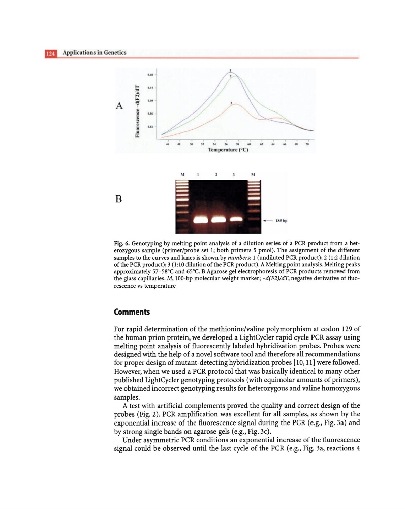
Fig. 6. Genotyping by melting point analysis of a dilution series of a PCR product from a heterozygous sample (primer/probe set 1; both primers 5 pmol). The assignment of the different
samples to the curves and lanes is shown by numbers: 1 (undiluted PCR product); 2 (1:2 dilution
of the PCR product); 3 (1:10 dilution of the PCR product). A Melting point analysis. Melting peaks
approximately 57-58°C and 65°C. B Agarose gel electrophoresis of PCR products removed from
the glass capillaries. M, 100-bp molecular weight marker; -d(F2}!dT, negative derivative of fluorescence vs temperature
Comments
For rapid determination of the methionine/valine polymorphism at codon 129 of
the human prion protein, we developed a LightCycler rapid cycle PCR assay using
melting point analysis of fluorescently labeled hybridization probes. Probes were
designed with the help of a novel software tool and therefore all recommendations
for proper design of mutant -detecting hybridization probes [10, III were followed.
However, when we used a PCR protocol that was basically identical to many other
published LightCycler genotyping protocols (with equimolar amounts of primers),
we obtained incorrect genotyping results for heterozygous and valine homozygous
samples.
A test with artificial complements proved the quality and correct design of the
probes (Fig. 2). PCR amplification was excellent for all samples, as shown by the
exponential increase of the fluorescence signal during the PCR (e.g., Fig. 3a) and
by strong single bands on agarose gels (e.g., Fig. 3c).
Under asymmetric PCR conditions an exponential increase of the fluorescence
signal could be observed until the last cycle of the PCR (e.g., Fig. 3a, reactions 4
A
B
I.,.
...
....
I
"
I
"
~
56
U
.;
. . . . .. .. ,.
Temperature (DC)
Fig. 6. Genotyping by melting point analysis of a dilution series of a PCR product from a heterozygous sample (primer/probe set 1; both primers 5 pmol). The assignment of the different
samples to the curves and lanes is shown by numbers: 1 (undiluted PCR product); 2 (1:2 dilution
of the PCR product); 3 (1:10 dilution of the PCR product). A Melting point analysis. Melting peaks
approximately 57-58°C and 65°C. B Agarose gel electrophoresis of PCR products removed from
the glass capillaries. M, 100-bp molecular weight marker; -d(F2}!dT, negative derivative of fluorescence vs temperature
Comments
For rapid determination of the methionine/valine polymorphism at codon 129 of
the human prion protein, we developed a LightCycler rapid cycle PCR assay using
melting point analysis of fluorescently labeled hybridization probes. Probes were
designed with the help of a novel software tool and therefore all recommendations
for proper design of mutant -detecting hybridization probes [10, III were followed.
However, when we used a PCR protocol that was basically identical to many other
published LightCycler genotyping protocols (with equimolar amounts of primers),
we obtained incorrect genotyping results for heterozygous and valine homozygous
samples.
A test with artificial complements proved the quality and correct design of the
probes (Fig. 2). PCR amplification was excellent for all samples, as shown by the
exponential increase of the fluorescence signal during the PCR (e.g., Fig. 3a) and
by strong single bands on agarose gels (e.g., Fig. 3c).
Under asymmetric PCR conditions an exponential increase of the fluorescence
signal could be observed until the last cycle of the PCR (e.g., Fig. 3a, reactions 4