

Eph Receptor Tyrosine Kinases and Their Ligands in Development 157
200112 -
112A
B
N
~==~==~-===~~--J
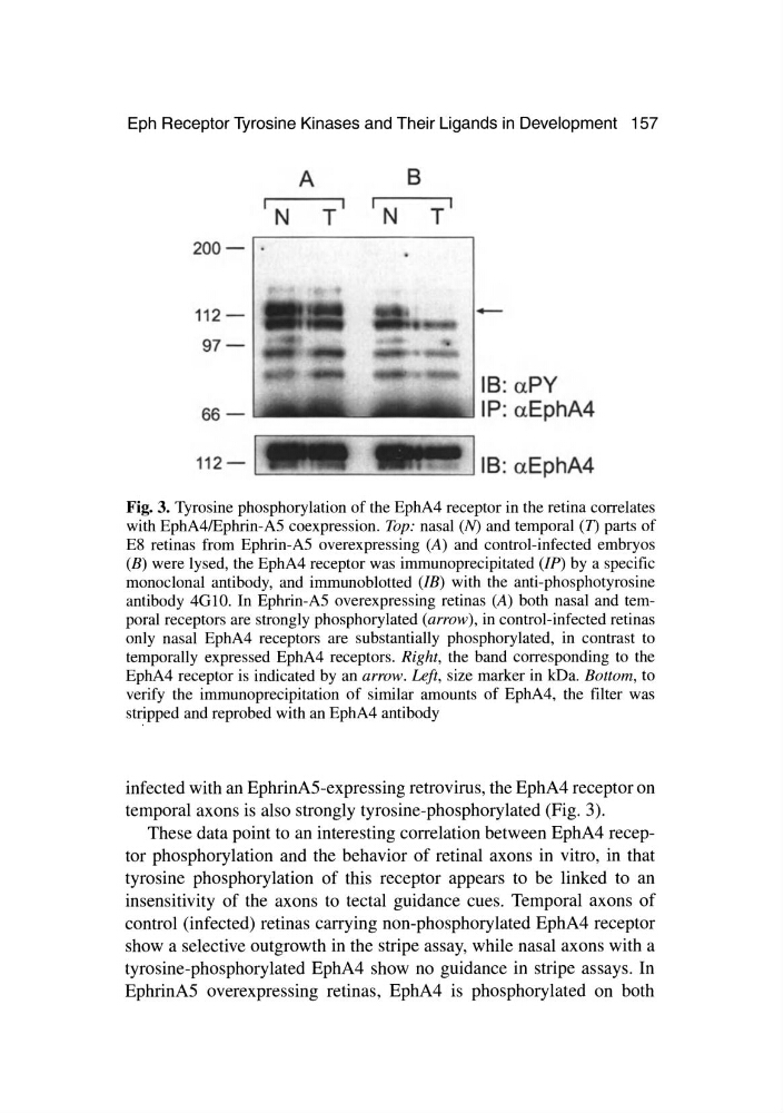
Fig. 3. Tyrosine phosphorylation of the EphA4 receptor in the retina correlates
with EphA4lEphrin-A5 coexpression. Top: nasal (N) and temporal (n parts of
E8 retinas from Ephrin-A5 overexpressing (A) and control-infected embryos
(B) were lysed, the EphA4 receptor was immunoprecipitated (IP) by a specific
monoclonal antibody, and immunoblotted (IB) with the anti-phosphotyrosine
antibody 4GlO. In Ephrin-A5 overexpressing retinas (A) both nasal and temporal receptors are strongly phosphorylated (arrow), in control-infected retinas
only nasal EphA4 receptors are substantially phosphorylated, in contrast to
temporally expressed EphA4 receptors. Right, the band corresponding to the
EphA4 receptor is indicated by an arrow. Left, size marker in kDa. Bottom, to
verify the immunoprecipitation of similar amounts of EphA4, the filter was
stripped and reprobed with an EphA4 antibody
infected with an EphrinAS-expressing retrovirus, the EphA4 receptor on
temporal axons is also strongly tyrosine-phosphorylated (Fig. 3).
These data point to an interesting correlation between EphA4 receptor phosphorylation and the behavior of retinal axons in vitro, in that
tyrosine phosphorylation of this receptor appears to be linked to an
insensitivity of the axons to tectal guidance cues. Temporal axons of
control (infected) retinas carrying non-phosphorylated EphA4 receptor
show a selective outgrowth in the stripe assay, while nasal axons with a
tyrosine-phosphorylated EphA4 show no guidance in stripe assays. In
EphrinAS overexpressing retinas, EphA4 is phosphorylated on both
200112 -
112A
B
N
~==~==~-===~~--J
Fig. 3. Tyrosine phosphorylation of the EphA4 receptor in the retina correlates
with EphA4lEphrin-A5 coexpression. Top: nasal (N) and temporal (n parts of
E8 retinas from Ephrin-A5 overexpressing (A) and control-infected embryos
(B) were lysed, the EphA4 receptor was immunoprecipitated (IP) by a specific
monoclonal antibody, and immunoblotted (IB) with the anti-phosphotyrosine
antibody 4GlO. In Ephrin-A5 overexpressing retinas (A) both nasal and temporal receptors are strongly phosphorylated (arrow), in control-infected retinas
only nasal EphA4 receptors are substantially phosphorylated, in contrast to
temporally expressed EphA4 receptors. Right, the band corresponding to the
EphA4 receptor is indicated by an arrow. Left, size marker in kDa. Bottom, to
verify the immunoprecipitation of similar amounts of EphA4, the filter was
stripped and reprobed with an EphA4 antibody
infected with an EphrinAS-expressing retrovirus, the EphA4 receptor on
temporal axons is also strongly tyrosine-phosphorylated (Fig. 3).
These data point to an interesting correlation between EphA4 receptor phosphorylation and the behavior of retinal axons in vitro, in that
tyrosine phosphorylation of this receptor appears to be linked to an
insensitivity of the axons to tectal guidance cues. Temporal axons of
control (infected) retinas carrying non-phosphorylated EphA4 receptor
show a selective outgrowth in the stripe assay, while nasal axons with a
tyrosine-phosphorylated EphA4 show no guidance in stripe assays. In
EphrinAS overexpressing retinas, EphA4 is phosphorylated on both