

156
U. Drescher
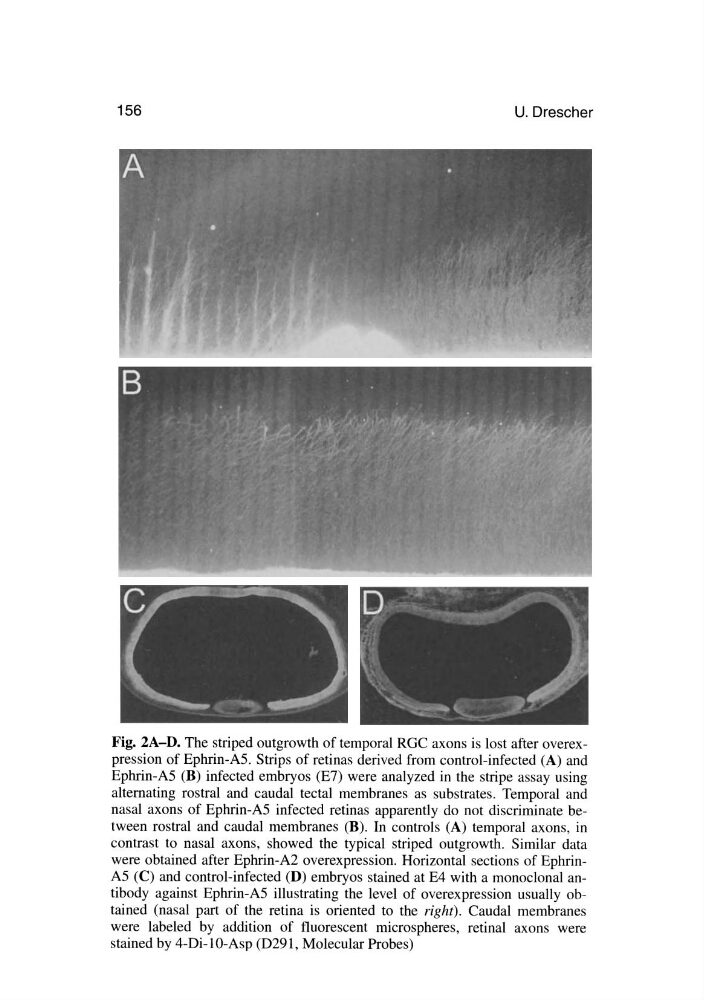
Fig. 2A-D. The striped outgrowth of temporal RGC axons is lost after overexpression of Ephrin-AS. Strips of retinas derived from control-infected (A) and
Ephrin-AS (B) infected embryos (E7) were analyzed in the stripe assay using
alternating rostral and caudal tectal membranes as substrates. Temporal and
nasal axons of Ephrin-AS infected retinas apparently do not discriminate between rostral and caudal membranes (B). In controls (A) temporal axons, in
contrast to nasal axons, showed the typical striped outgrowth. Similar data
were obtained after Ephrin-A2 overexpression. Horizontal sections of EphrinAS (C) and control-infected (D) embryos stained at E4 with a monoclonal antibody against Ephrin-AS illustrating the level of overexpression usually obtained (nasal part of the retina is oriented to the right). Caudal membranes
were labeled by addition of fluorescent microspheres, retinal axons were
stained by 4-Di-1O-Asp (D291, Molecular Probes)
U. Drescher
Fig. 2A-D. The striped outgrowth of temporal RGC axons is lost after overexpression of Ephrin-AS. Strips of retinas derived from control-infected (A) and
Ephrin-AS (B) infected embryos (E7) were analyzed in the stripe assay using
alternating rostral and caudal tectal membranes as substrates. Temporal and
nasal axons of Ephrin-AS infected retinas apparently do not discriminate between rostral and caudal membranes (B). In controls (A) temporal axons, in
contrast to nasal axons, showed the typical striped outgrowth. Similar data
were obtained after Ephrin-A2 overexpression. Horizontal sections of EphrinAS (C) and control-infected (D) embryos stained at E4 with a monoclonal antibody against Ephrin-AS illustrating the level of overexpression usually obtained (nasal part of the retina is oriented to the right). Caudal membranes
were labeled by addition of fluorescent microspheres, retinal axons were
stained by 4-Di-1O-Asp (D291, Molecular Probes)