

Eph Receptor Tyrosine Kinases and Their Ligands in Development 155
retina
tectum
temp.
nasal
rostral
caudal
EphA3
EphA4, EphAS 1,--------,
Ephrin-A2
~ Ephrin-A2
Ephrin-AS
~ Ephrin-AS
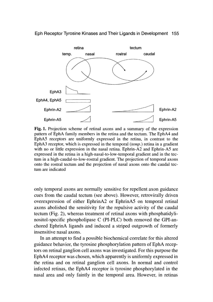
Fig. 1. Projection scheme of retinal axons and a summary of the expression
pattern of EphA family members in the retina and the tectum. The EphA4 and
EphA5 receptors are uniformly expressed in the retina, in contrast to the
EphA3 receptor, which is expressed in the temporal (temp.) retina in a gradient
with no or little expression in the nasal retina. Ephrin-A2 and Ephrin-A5 are
expressed in the retina in a high-nasal-to-low-temporal gradient and in the tectum in a high-caudal-to-low-rostral gradient. The projection of temporal axons
onto the rostral tectum and the projection of nasal axons onto the caudal tectum are indicated
only temporal axons are normally sensitive for repellent axon guidance
cues from the caudal tectum (see above). However, retrovirally driven
overexpression of either EphrinA2 or EphrinAS on temporal retinal
axons abolished the sensitivity for the repulsive activity of the caudal
tectum (Fig. 2), whereas treatment of retinal axons with phosphatidylinositol-specific phospholipase C (PI-PLC) both removed the GPI-anchored EphrinA ligands and induced a striped outgrowth of formerly
insensitive nasal axons.
In an attempt to find a possible biochemical correlate for this altered
guidance behavior, the tyrosine phosphorylation pattern of EphA receptors on retinal ganglion cell axons was investigated. For this purpose the
EphA4 receptor was chosen, which apparently is uniformly expressed in
the retina and on retinal ganglion cell axons. In normal and control
infected retinas, the EphA4 receptor is tyrosine phosphorylated in the
nasal area and only faintly in the temporal area. However, in retinas
retina
tectum
temp.
nasal
rostral
caudal
EphA3
EphA4, EphAS 1,--------,
Ephrin-A2
~ Ephrin-A2
Ephrin-AS
~ Ephrin-AS
Fig. 1. Projection scheme of retinal axons and a summary of the expression
pattern of EphA family members in the retina and the tectum. The EphA4 and
EphA5 receptors are uniformly expressed in the retina, in contrast to the
EphA3 receptor, which is expressed in the temporal (temp.) retina in a gradient
with no or little expression in the nasal retina. Ephrin-A2 and Ephrin-A5 are
expressed in the retina in a high-nasal-to-low-temporal gradient and in the tectum in a high-caudal-to-low-rostral gradient. The projection of temporal axons
onto the rostral tectum and the projection of nasal axons onto the caudal tectum are indicated
only temporal axons are normally sensitive for repellent axon guidance
cues from the caudal tectum (see above). However, retrovirally driven
overexpression of either EphrinA2 or EphrinAS on temporal retinal
axons abolished the sensitivity for the repulsive activity of the caudal
tectum (Fig. 2), whereas treatment of retinal axons with phosphatidylinositol-specific phospholipase C (PI-PLC) both removed the GPI-anchored EphrinA ligands and induced a striped outgrowth of formerly
insensitive nasal axons.
In an attempt to find a possible biochemical correlate for this altered
guidance behavior, the tyrosine phosphorylation pattern of EphA receptors on retinal ganglion cell axons was investigated. For this purpose the
EphA4 receptor was chosen, which apparently is uniformly expressed in
the retina and on retinal ganglion cell axons. In normal and control
infected retinas, the EphA4 receptor is tyrosine phosphorylated in the
nasal area and only faintly in the temporal area. However, in retinas