

158
T
N
removal of
~s
T
relroviral overexpression
of~ns
T
U. Drescher
N
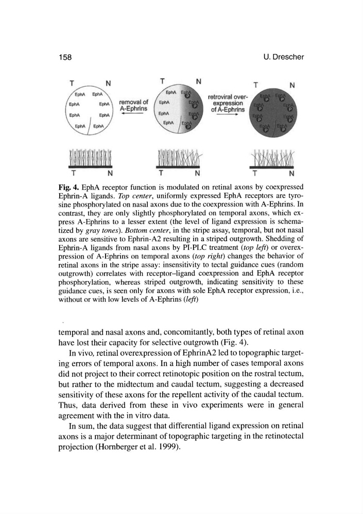
Fig. 4. EphA receptor function is modulated on retinal axons by coexpressed
Ephrin-A ligands. Top center, uniformly expressed EphA receptors are tyrosine phosphorylated on nasal axons due to the coexpression with A-Ephrins. In
contrast, they are only slightly phosphorylated on temporal axons, which express A-Ephrins to a lesser extent (the level of ligand expression is schematized by gray tones). Bottom center, in the stripe assay, temporal, but not nasal
axons are sensitive to Ephrin-A2 resulting in a striped outgrowth. Shedding of
Ephrin-A ligands from nasal axons by PI-PLC treatment (top left) or overexpression of A-Ephrins on temporal axons (top right) changes the behavior of
retinal axons in the stripe assay: insensitivity to tectal guidance cues (random
outgrowth) correlates with receptor-ligand coexpression and EphA receptor
phosphorylation, whereas striped outgrowth, indicating sensitivity to these
guidance cues, is seen only for axons with sole EphA receptor expression, i.e.,
without or with low levels of A-Ephrins (left)
temporal and nasal axons and, concomitantly, both types of retinal axon
have lost their capacity for selective outgrowth (Fig. 4).
In vivo, retinal overexpression of EphrinA2 led to topographic targeting errors of temporal axons. In a high number of cases temporal axons
did not project to their correct retinotopic position on the rostral tectum,
but rather to the midtectum and caudal tectum, suggesting a decreased
sensitivity of these axons for the repellent activity of the caudal tectum.
Thus, data derived from these in vivo experiments were in general
agreement with the in vitro data.
In sum, the data suggest that differential ligand expression on retinal
axons is a major determinant of topographic targeting in the retinotectal
projection (Hornberger et al. 1999).
T
N
removal of
~s
T
relroviral overexpression
of~ns
T
U. Drescher
N
Fig. 4. EphA receptor function is modulated on retinal axons by coexpressed
Ephrin-A ligands. Top center, uniformly expressed EphA receptors are tyrosine phosphorylated on nasal axons due to the coexpression with A-Ephrins. In
contrast, they are only slightly phosphorylated on temporal axons, which express A-Ephrins to a lesser extent (the level of ligand expression is schematized by gray tones). Bottom center, in the stripe assay, temporal, but not nasal
axons are sensitive to Ephrin-A2 resulting in a striped outgrowth. Shedding of
Ephrin-A ligands from nasal axons by PI-PLC treatment (top left) or overexpression of A-Ephrins on temporal axons (top right) changes the behavior of
retinal axons in the stripe assay: insensitivity to tectal guidance cues (random
outgrowth) correlates with receptor-ligand coexpression and EphA receptor
phosphorylation, whereas striped outgrowth, indicating sensitivity to these
guidance cues, is seen only for axons with sole EphA receptor expression, i.e.,
without or with low levels of A-Ephrins (left)
temporal and nasal axons and, concomitantly, both types of retinal axon
have lost their capacity for selective outgrowth (Fig. 4).
In vivo, retinal overexpression of EphrinA2 led to topographic targeting errors of temporal axons. In a high number of cases temporal axons
did not project to their correct retinotopic position on the rostral tectum,
but rather to the midtectum and caudal tectum, suggesting a decreased
sensitivity of these axons for the repellent activity of the caudal tectum.
Thus, data derived from these in vivo experiments were in general
agreement with the in vitro data.
In sum, the data suggest that differential ligand expression on retinal
axons is a major determinant of topographic targeting in the retinotectal
projection (Hornberger et al. 1999).