

12
reducing ends releasing xylooligosaccharides; and β-xylosidases, which cleaves the
xylobiose and xylooligosaccharides to release xylose. In addition, the enzymes
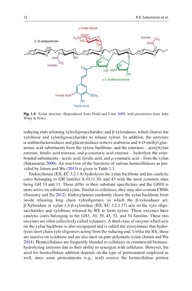
α-arabinofuranosidases and glucuronidases remove arabinose and 4-O-methyl glucuronic acid substituents from the xylose backbone, and the esterases – acetylxylan
esterase, ferulic acid esterase, and ρ-coumaric acid esterase – hydrolyze the esterbonded substituents – acetic acid, ferulic acid, and ρ-coumaric acid – from the xylan
(Sukumaran 2009). An overview of the functions of various hemicellulases as provided by Juturu and Wu (2013) is given in Table 1.1.
Endoxylanase (EX, EC 3.2.1.8) hydrolyzes the xylan backbone and has catalytic
cores belonging to GH families 8,10,11,30, and 43 with the most common ones
being GH 10 and 11. These differ in their substrate specificities and the GH10 is
more active on substituted xylan. Similar to cellulases, they may also contain CBMs
(Sweeney and Xu 2012). Endoxylanases randomly cleave the xylan backbone from
inside releasing long chain xylooligomers on which the β-xylosidases act.
β-Xylosidase or xylan-1,4-β-xylosidase (BX, EC 3.2.1.37) acts on the xylo oligosaccharides and xylobiose released by BX to form xylose. These enzymes have
catalytic cores belonging to the GH3, 30, 39, 43, 52, and 54 families. These two
enzymes are often collectively called xylanases. A third class of enzyme which acts
on the xylan backbone is also recognized and is called the exoxylanase that hydrolyzes short chain xylo oligomers acting from the reducing end. Unlike the BX, these
are inactive on xylobiose and are also inert on pure polymeric xylan (Juturu and Wu
2014). Hemicellulases are frequently blended to cellulases in commercial biomasshydrolyzing enzymes due to their ability to synergize with cellulases. However, the
need for hemicellulase addition depends on the type of pretreatment employed as
well, since some pretreatments (e.g., acid) remove the hemicellulose portion
Fig. 1.4 Xylan structure (Reproduced from Dodd and Cann 2009, with permission from John
Wiley & Sons)
R.K. Sukumaran et al.
reducing ends releasing xylooligosaccharides; and β-xylosidases, which cleaves the
xylobiose and xylooligosaccharides to release xylose. In addition, the enzymes
α-arabinofuranosidases and glucuronidases remove arabinose and 4-O-methyl glucuronic acid substituents from the xylose backbone, and the esterases – acetylxylan
esterase, ferulic acid esterase, and ρ-coumaric acid esterase – hydrolyze the esterbonded substituents – acetic acid, ferulic acid, and ρ-coumaric acid – from the xylan
(Sukumaran 2009). An overview of the functions of various hemicellulases as provided by Juturu and Wu (2013) is given in Table 1.1.
Endoxylanase (EX, EC 3.2.1.8) hydrolyzes the xylan backbone and has catalytic
cores belonging to GH families 8,10,11,30, and 43 with the most common ones
being GH 10 and 11. These differ in their substrate specificities and the GH10 is
more active on substituted xylan. Similar to cellulases, they may also contain CBMs
(Sweeney and Xu 2012). Endoxylanases randomly cleave the xylan backbone from
inside releasing long chain xylooligomers on which the β-xylosidases act.
β-Xylosidase or xylan-1,4-β-xylosidase (BX, EC 3.2.1.37) acts on the xylo oligosaccharides and xylobiose released by BX to form xylose. These enzymes have
catalytic cores belonging to the GH3, 30, 39, 43, 52, and 54 families. These two
enzymes are often collectively called xylanases. A third class of enzyme which acts
on the xylan backbone is also recognized and is called the exoxylanase that hydrolyzes short chain xylo oligomers acting from the reducing end. Unlike the BX, these
are inactive on xylobiose and are also inert on pure polymeric xylan (Juturu and Wu
2014). Hemicellulases are frequently blended to cellulases in commercial biomasshydrolyzing enzymes due to their ability to synergize with cellulases. However, the
need for hemicellulase addition depends on the type of pretreatment employed as
well, since some pretreatments (e.g., acid) remove the hemicellulose portion
Fig. 1.4 Xylan structure (Reproduced from Dodd and Cann 2009, with permission from John
Wiley & Sons)
R.K. Sukumaran et al.