

296
5'
~
r;:
j(KM)
c
8. 400J
11
lOOJ
'"
1CMK1
ICMKI
·ICMKI
·iiI
iii
100
150
100
LIO
JOO
Time Is)
-
100 o
H. TSCHESCHE and M. FARR
1
0.5
1
1.0
1.5
2.0
c(MM P) I ~M l
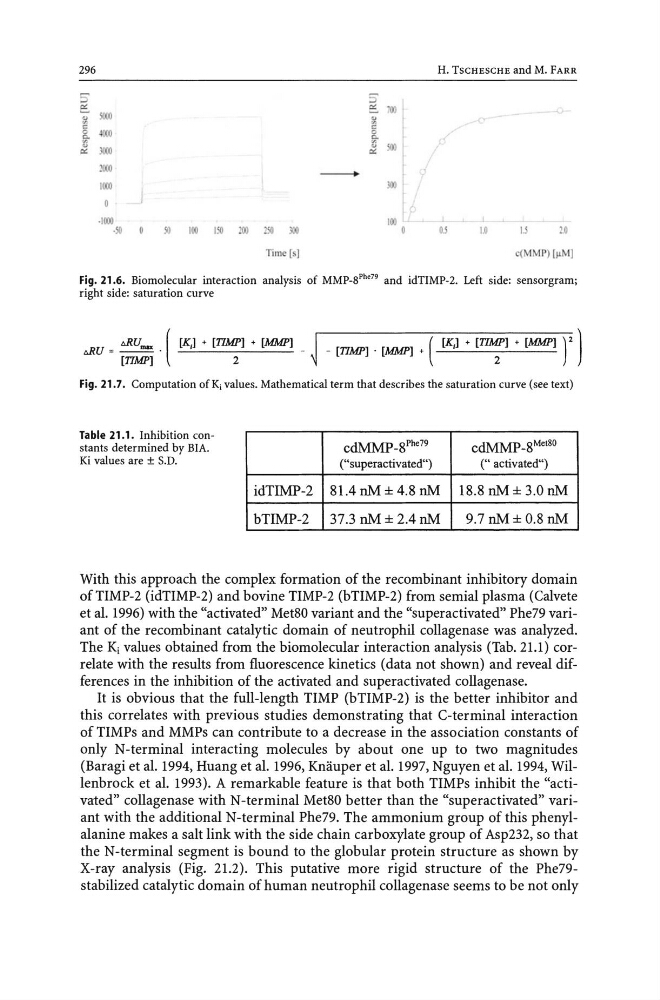
Fig. 21.6. Biomolecular interaction analysis of MMP_SPhe79 and idTIMP-2. Left side: sensorgram;
right side: saturation curve
t>RU = ~~ . ( [K,] + [~] + [MMP] _ ~ _ [TIMP] . [MMP] + ( [K,] + [TI~] + [MMP] ) 2 )
Fig. 21.7. Computation ofKj values. Mathematical term that describes the saturation curve (see text)
Table 21.1. Inhibition constants determined by BIA.
Ki values are ± S.D.
idTIMP-2
bTIMP-2
cdMMP_8Phe79
("superactivated")
81.4 nM ± 4.8 nM
37.3 nM ± 2.4 nM
cdMMP_8Met80
(" activated")
18.8 nM ± 3.0 nM
9.7 nM ± 0.8 nM
With this approach the complex formation of the recombinant inhibitory domain
ofTIMP-2 (idTIMP-2) and bovine TIMP-2 (bTIMP-2) from semial plasma (Calvete
et al. 1996) with the "activated" Met80 variant and the "superactivated" Phe79 variant of the recombinant catalytic domain of neutrophil collagenase was analyzed.
The Ki values obtained from the biomolecular interaction analysis (Tab. 21.1) correlate with the results from fluorescence kinetics (data not shown) and reveal differences in the inhibition of the activated and superactivated collagenase.
It is obvious that the full-length TIMP (bTIMP-2) is the better inhibitor and
this correlates with previous studies demonstrating that C-terminal interaction
of TIMPs and MMPs can contribute to a decrease in the association constants of
only N-terminal interacting molecules by about one up to two magnitudes
(Baragi et al. 1994, Huang et al. 1996, Knauper et al. 1997, Nguyen et al. 1994, Willenbrock et al. 1993). A remarkable feature is that both TIMPs inhibit the "activated" collagenase with N-terminal Met80 better than the "superactivated" variant with the additional N-terminal Phe79. The ammonium group of this phenylalanine makes a salt link with the side chain carboxylate group of Asp232, so that
the N-terminal segment is bound to the globular protein structure as shown by
X-ray analysis (Fig. 21.2). This putative more rigid structure of the Phe79stabilized catalytic domain of human neutrophil collagenase seems to be not only
5'
~
r;:
j(KM)
c
8. 400J
11
lOOJ
'"
1CMK1
ICMKI
·ICMKI
·iiI
iii
100
150
100
LIO
JOO
Time Is)
-
100 o
H. TSCHESCHE and M. FARR
1
0.5
1
1.0
1.5
2.0
c(MM P) I ~M l
Fig. 21.6. Biomolecular interaction analysis of MMP_SPhe79 and idTIMP-2. Left side: sensorgram;
right side: saturation curve
t>RU = ~~ . ( [K,] + [~] + [MMP] _ ~ _ [TIMP] . [MMP] + ( [K,] + [TI~] + [MMP] ) 2 )
Fig. 21.7. Computation ofKj values. Mathematical term that describes the saturation curve (see text)
Table 21.1. Inhibition constants determined by BIA.
Ki values are ± S.D.
idTIMP-2
bTIMP-2
cdMMP_8Phe79
("superactivated")
81.4 nM ± 4.8 nM
37.3 nM ± 2.4 nM
cdMMP_8Met80
(" activated")
18.8 nM ± 3.0 nM
9.7 nM ± 0.8 nM
With this approach the complex formation of the recombinant inhibitory domain
ofTIMP-2 (idTIMP-2) and bovine TIMP-2 (bTIMP-2) from semial plasma (Calvete
et al. 1996) with the "activated" Met80 variant and the "superactivated" Phe79 variant of the recombinant catalytic domain of neutrophil collagenase was analyzed.
The Ki values obtained from the biomolecular interaction analysis (Tab. 21.1) correlate with the results from fluorescence kinetics (data not shown) and reveal differences in the inhibition of the activated and superactivated collagenase.
It is obvious that the full-length TIMP (bTIMP-2) is the better inhibitor and
this correlates with previous studies demonstrating that C-terminal interaction
of TIMPs and MMPs can contribute to a decrease in the association constants of
only N-terminal interacting molecules by about one up to two magnitudes
(Baragi et al. 1994, Huang et al. 1996, Knauper et al. 1997, Nguyen et al. 1994, Willenbrock et al. 1993). A remarkable feature is that both TIMPs inhibit the "activated" collagenase with N-terminal Met80 better than the "superactivated" variant with the additional N-terminal Phe79. The ammonium group of this phenylalanine makes a salt link with the side chain carboxylate group of Asp232, so that
the N-terminal segment is bound to the globular protein structure as shown by
X-ray analysis (Fig. 21.2). This putative more rigid structure of the Phe79stabilized catalytic domain of human neutrophil collagenase seems to be not only