

294
H. TSCHESCHE and M. FARR
c
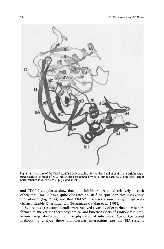
Fig. 21.4. Structure of the TIMP-2/MTl-MMP complex (Fernandez-Catalan et al. 1998). Bright structure: catalytic domain of MTl-MMP; dark structure: bovine TIMP-2; dark balls: zinc ions; bright
balls: calcium ions; h: helix; s: p-pleated sheet
and TIMP-2 complexes show that both inhibitors are tilted relatively to each
other, that TIMP-2 has a quite elongated sA-sB j3-hairpin loop that rises above
the j3-barrel (Fig. 21.4), and that TIMP-2 possesses a much longer negatively
charged flexible C-terminal tail (Fernandez-Catalan et al. 1998).
Before these structural details were resolved a variety of experiments was performed to analyze the thermodynamical and kinetic aspects ofTIMP/MMP interaction using labelled synthetic or physiological substrates. One of the recent
methods to analyze these biomolecular interactions are the BIA-systems
H. TSCHESCHE and M. FARR
c
Fig. 21.4. Structure of the TIMP-2/MTl-MMP complex (Fernandez-Catalan et al. 1998). Bright structure: catalytic domain of MTl-MMP; dark structure: bovine TIMP-2; dark balls: zinc ions; bright
balls: calcium ions; h: helix; s: p-pleated sheet
and TIMP-2 complexes show that both inhibitors are tilted relatively to each
other, that TIMP-2 has a quite elongated sA-sB j3-hairpin loop that rises above
the j3-barrel (Fig. 21.4), and that TIMP-2 possesses a much longer negatively
charged flexible C-terminal tail (Fernandez-Catalan et al. 1998).
Before these structural details were resolved a variety of experiments was performed to analyze the thermodynamical and kinetic aspects ofTIMP/MMP interaction using labelled synthetic or physiological substrates. One of the recent
methods to analyze these biomolecular interactions are the BIA-systems