

Epitope Mapping for the Monoclonal Antibody that Inhibits Intramolecular Flavin
153
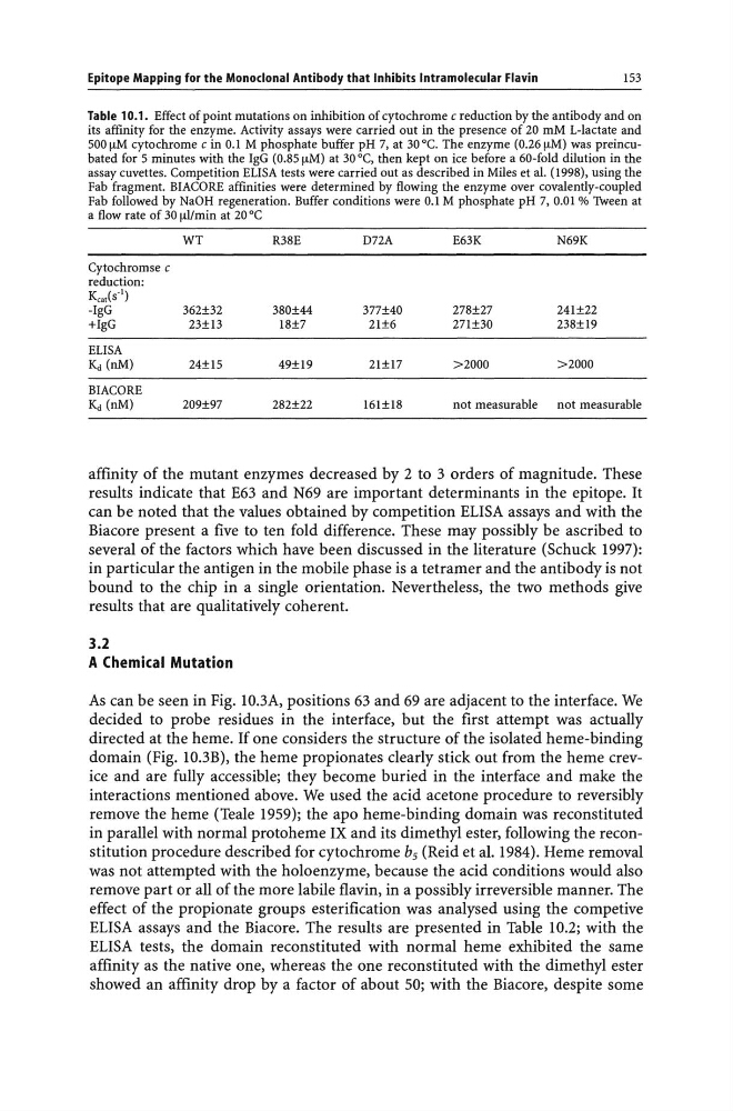
Table 10.1. Effect of point mutations on inhibition of cytochrome c reduction by the antibody and on
its affinity for the enzyme. Activity assays were carried out in the presence of 20 mM L-Iactate and
500 [LM cytochrome c in 0.1 M phosphate buffer pH 7, at 30°C. The enzyme (0.26 [LM) was preincubated for 5 minutes with the IgG (0.85 [LM) at 30°C, then kept on ice before a 60-fold dilution in the
assay cuvettes. Competition ELISA tests were carried out as described in Miles et al. (1998), using the
Fab fragment. BIACORE affinities were determined by flowing the enzyme over covalently-coupled
Fab followed by NaOH regeneration. Buffer conditions were 0.1 M phosphate pH 7, 0.01 % Tween at
a flow rate of 30 [LI/min at 20°C
WT
Cytochromse c
reduction:
Keat(s'!)
-IgG
362±32
+IgG
23±13
ELISA
Kd (nM)
24±15
BIACORE
Kd (nM)
209±97
R38E
380±44
18±7
49±19
282±22
DnA
377±40
21±6
21±17
161±18
E63K
N69K
278±27
241±22
271±30
238±19
>2000
>2000
not measurable
not measurable
affinity of the mutant enzymes decreased by 2 to 3 orders of magnitude. These
results indicate that E63 and N69 are important determinants in the epitope. It
can be noted that the values obtained by competition ELISA assays and with the
Biacore present a five to ten fold difference. These may possibly be ascribed to
several of the factors which have been discussed in the literature (Schuck 1997):
in particular the antigen in the mobile phase is a tetramer and the antibody is not
bound to the chip in a single orientation. Nevertheless, the two methods give
results that are qualitatively coherent.
3.2
A Chemical Mutation
As can be seen in Fig. 1O.3A, positions 63 and 69 are adjacent to the interface. We
decided to probe residues in the interface, but the first attempt was actually
directed at the heme. If one considers the structure of the isolated heme-binding
domain (Fig. 10.3B), the heme propionates clearly stick out from the heme crevice and are fully accessible; they become buried in the interface and make the
interactions mentioned above. We used the acid acetone procedure to reversibly
remove the heme (Teale 1959); the apo heme-binding domain was reconstituted
in parallel with normal protoheme IX and its dimethyl ester, following the reconstitution procedure described for cytochrome bs (Reid et al. 1984). Heme removal
was not attempted with the holoenzyme, because the acid conditions would also
remove part or all of the more labile flavin, in a possibly irreversible manner. The
effect of the propionate groups esterification was analysed using the competive
ELISA assays and the Biacore. The results are presented in Table 10.2; with the
ELISA tests, the domain reconstituted with normal heme exhibited the same
affinity as the native one, whereas the one reconstituted with the dimethyl ester
showed an affinity drop by a factor of about 50; with the Biacore, despite some
153
Table 10.1. Effect of point mutations on inhibition of cytochrome c reduction by the antibody and on
its affinity for the enzyme. Activity assays were carried out in the presence of 20 mM L-Iactate and
500 [LM cytochrome c in 0.1 M phosphate buffer pH 7, at 30°C. The enzyme (0.26 [LM) was preincubated for 5 minutes with the IgG (0.85 [LM) at 30°C, then kept on ice before a 60-fold dilution in the
assay cuvettes. Competition ELISA tests were carried out as described in Miles et al. (1998), using the
Fab fragment. BIACORE affinities were determined by flowing the enzyme over covalently-coupled
Fab followed by NaOH regeneration. Buffer conditions were 0.1 M phosphate pH 7, 0.01 % Tween at
a flow rate of 30 [LI/min at 20°C
WT
Cytochromse c
reduction:
Keat(s'!)
-IgG
362±32
+IgG
23±13
ELISA
Kd (nM)
24±15
BIACORE
Kd (nM)
209±97
R38E
380±44
18±7
49±19
282±22
DnA
377±40
21±6
21±17
161±18
E63K
N69K
278±27
241±22
271±30
238±19
>2000
>2000
not measurable
not measurable
affinity of the mutant enzymes decreased by 2 to 3 orders of magnitude. These
results indicate that E63 and N69 are important determinants in the epitope. It
can be noted that the values obtained by competition ELISA assays and with the
Biacore present a five to ten fold difference. These may possibly be ascribed to
several of the factors which have been discussed in the literature (Schuck 1997):
in particular the antigen in the mobile phase is a tetramer and the antibody is not
bound to the chip in a single orientation. Nevertheless, the two methods give
results that are qualitatively coherent.
3.2
A Chemical Mutation
As can be seen in Fig. 1O.3A, positions 63 and 69 are adjacent to the interface. We
decided to probe residues in the interface, but the first attempt was actually
directed at the heme. If one considers the structure of the isolated heme-binding
domain (Fig. 10.3B), the heme propionates clearly stick out from the heme crevice and are fully accessible; they become buried in the interface and make the
interactions mentioned above. We used the acid acetone procedure to reversibly
remove the heme (Teale 1959); the apo heme-binding domain was reconstituted
in parallel with normal protoheme IX and its dimethyl ester, following the reconstitution procedure described for cytochrome bs (Reid et al. 1984). Heme removal
was not attempted with the holoenzyme, because the acid conditions would also
remove part or all of the more labile flavin, in a possibly irreversible manner. The
effect of the propionate groups esterification was analysed using the competive
ELISA assays and the Biacore. The results are presented in Table 10.2; with the
ELISA tests, the domain reconstituted with normal heme exhibited the same
affinity as the native one, whereas the one reconstituted with the dimethyl ester
showed an affinity drop by a factor of about 50; with the Biacore, despite some