

152
3
Results
3.1
K. H. DIEP LE et al.
Site-Directed Mutagenesis of the Heme-Binding Domain in Flavocytochrome bz
For a start we chose to mutate charged residues or polar ones. Four mutations
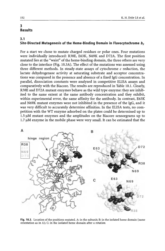
were individually introduced: R38E, E63K, N69K and DnA. The first position
mutated lies at the "waist" of the heme-binding domain, the three others are very
close to the interface (Fig. lO.3A). The effect of the mutations was assessed using
three different methods. In steady-state assays of cytochrome c reduction, the
lactate dehydrogenase activity at saturating substrate and acceptor concentrations was compared in the presence and absence of a fixed IgG concentration. In
parallel, dissociation constants were analyzed in competitive ELISA assays and
comparatively with the Biacore. The results are reproduced in Table 10.1. Clearly,
R38E and DnA mutant enzymes behave as the wild type enzyme: they are inhibited to the same extent at the same antibody concentration and they exhibit,
within experimental error, the same affinity for the antibody. In contrast, E63K
and N69K mutant enzymes were not inhibited in the presence of the IgG, and it
was very difficult to accurately determine affinities. In the ELISA tests, no competition with the WT enzyme adsorbed on the plates could be determined up to
1.5 !-tM mutant enzymes and the amplitudes on the Biacore sensorgrams up to
1.7 !-tM enzyme in the mobile phase were very small. It can be estimated that the
A
c
E63
N69
Fig. 10.3. Location of the positions mutated. A: in the subunit; B: in the isolated heme domain (same
orientation as in Al; C: in the isolated heme domain after a rotation
3
Results
3.1
K. H. DIEP LE et al.
Site-Directed Mutagenesis of the Heme-Binding Domain in Flavocytochrome bz
For a start we chose to mutate charged residues or polar ones. Four mutations
were individually introduced: R38E, E63K, N69K and DnA. The first position
mutated lies at the "waist" of the heme-binding domain, the three others are very
close to the interface (Fig. lO.3A). The effect of the mutations was assessed using
three different methods. In steady-state assays of cytochrome c reduction, the
lactate dehydrogenase activity at saturating substrate and acceptor concentrations was compared in the presence and absence of a fixed IgG concentration. In
parallel, dissociation constants were analyzed in competitive ELISA assays and
comparatively with the Biacore. The results are reproduced in Table 10.1. Clearly,
R38E and DnA mutant enzymes behave as the wild type enzyme: they are inhibited to the same extent at the same antibody concentration and they exhibit,
within experimental error, the same affinity for the antibody. In contrast, E63K
and N69K mutant enzymes were not inhibited in the presence of the IgG, and it
was very difficult to accurately determine affinities. In the ELISA tests, no competition with the WT enzyme adsorbed on the plates could be determined up to
1.5 !-tM mutant enzymes and the amplitudes on the Biacore sensorgrams up to
1.7 !-tM enzyme in the mobile phase were very small. It can be estimated that the
A
c
E63
N69
Fig. 10.3. Location of the positions mutated. A: in the subunit; B: in the isolated heme domain (same
orientation as in Al; C: in the isolated heme domain after a rotation