

Epitope Mapping for the Monoclonal Antibody that Inhibits Intramolecular Flavin
151
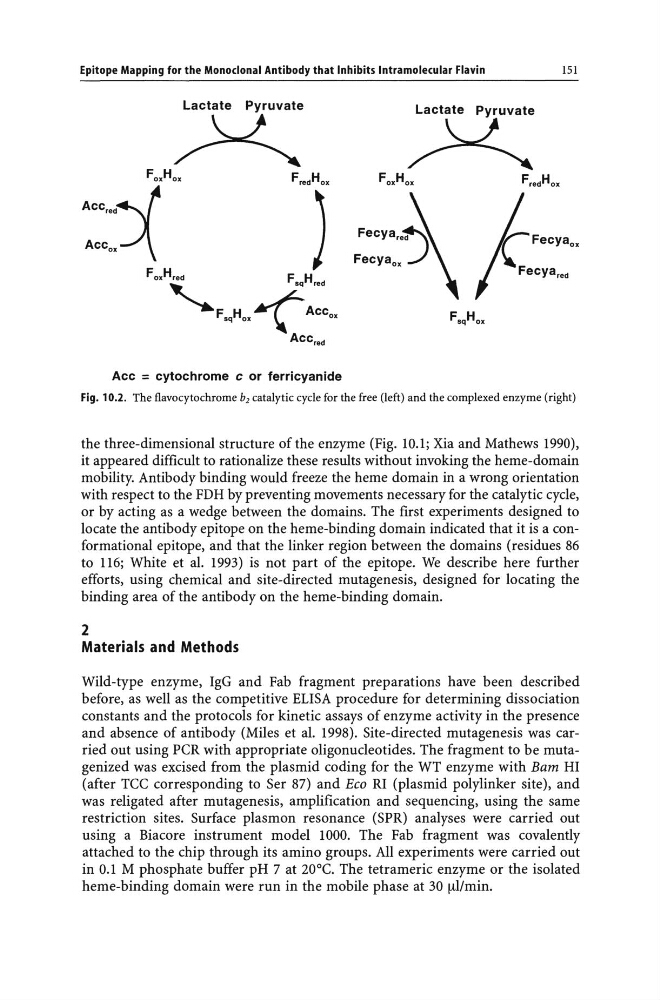
Acc = cytochrome c or ferricyanide
Fig. 10.2. The flavocytochrome b2 catalytic cycle for the free (left) and the complexed enzyme (right)
the three-dimensional structure of the enzyme (Fig. 10.1; Xia and Mathews 1990),
it appeared difficult to rationalize these results without invoking the heme-domain
mobility. Antibody binding would freeze the heme domain in a wrong orientation
with respect to the FDH by preventing movements necessary for the catalytic cycle,
or by acting as a wedge between the domains. The first experiments designed to
locate the antibody epitope on the heme-binding domain indicated that it is a conformational epitope, and that the linker region between the domains (residues 86
to 116; White et al. 1993) is not part of the epitope. We describe here further
efforts, using chemical and site-directed mutagenesis, designed for locating the
binding area of the antibody on the heme-binding domain.
2
Materials and Methods
Wild-type enzyme, IgG and Fab fragment preparations have been described
before, as well as the competitive ELISA procedure for determining dissociation
constants and the protocols for kinetic assays of enzyme activity in the presence
and absence of antibody (Miles et al. 1998). Site-directed mutagenesis was carried out using peR with appropriate oligonucleotides. The fragment to be mutagenized was excised from the plasmid coding for the WT enzyme with Bam HI
(after Tee corresponding to Ser 87) and Eeo RI (plasmid polylinker site), and
was religated after mutagenesis, amplification and sequencing, using the same
restriction sites. Surface plasmon resonance (SPR) analyses were carried out
using a Biacore instrument model 1000. The Fab fragment was covalently
attached to the chip through its amino groups. All experiments were carried out
in 0.1 M phosphate buffer pH 7 at 20°e. The tetrameric enzyme or the isolated
heme-binding domain were run in the mobile phase at 30 [ll/min.
151
Acc = cytochrome c or ferricyanide
Fig. 10.2. The flavocytochrome b2 catalytic cycle for the free (left) and the complexed enzyme (right)
the three-dimensional structure of the enzyme (Fig. 10.1; Xia and Mathews 1990),
it appeared difficult to rationalize these results without invoking the heme-domain
mobility. Antibody binding would freeze the heme domain in a wrong orientation
with respect to the FDH by preventing movements necessary for the catalytic cycle,
or by acting as a wedge between the domains. The first experiments designed to
locate the antibody epitope on the heme-binding domain indicated that it is a conformational epitope, and that the linker region between the domains (residues 86
to 116; White et al. 1993) is not part of the epitope. We describe here further
efforts, using chemical and site-directed mutagenesis, designed for locating the
binding area of the antibody on the heme-binding domain.
2
Materials and Methods
Wild-type enzyme, IgG and Fab fragment preparations have been described
before, as well as the competitive ELISA procedure for determining dissociation
constants and the protocols for kinetic assays of enzyme activity in the presence
and absence of antibody (Miles et al. 1998). Site-directed mutagenesis was carried out using peR with appropriate oligonucleotides. The fragment to be mutagenized was excised from the plasmid coding for the WT enzyme with Bam HI
(after Tee corresponding to Ser 87) and Eeo RI (plasmid polylinker site), and
was religated after mutagenesis, amplification and sequencing, using the same
restriction sites. Surface plasmon resonance (SPR) analyses were carried out
using a Biacore instrument model 1000. The Fab fragment was covalently
attached to the chip through its amino groups. All experiments were carried out
in 0.1 M phosphate buffer pH 7 at 20°e. The tetrameric enzyme or the isolated
heme-binding domain were run in the mobile phase at 30 [ll/min.