

150
Flavodehydrogenase
(FDH)
~--------~,,~----------/
K. H. DIEP LE et al.
Heme b 2 domain
r-----~I\~------~ ,
region
FMN
Heme propionates
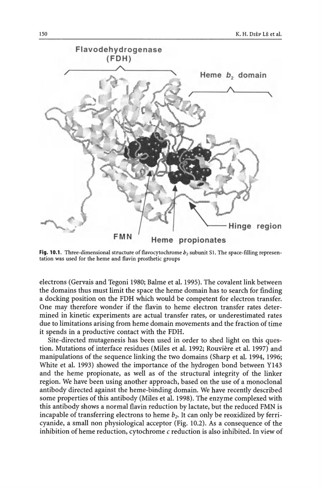
Fig. 10.1. Three-dimensional structure offlavocytochrome b2 subunit S1. The space-filling representation was used for the heme and flavin prosthetic groups
electrons (Gervais and Tegoni 1980; Balme et al. 1995). The covalent link between
the domains thus must limit the space the heme domain has to search for finding
a docking position on the FDH which would be competent for electron transfer.
One may therefore wonder if the flavin to heme electron transfer rates determined in kinetic experiments are actual transfer rates, or underestimated rates
due to limitations arising from heme domain movements and the fraction of time
it spends in a productive contact with the FDH.
Site-directed mutagenesis has been used in order to shed light on this question. Mutations of interface residues (Miles et al. 1992; Rouviere et al. 1997) and
manipulations of the sequence linking the two domains (Sharp et al. 1994, 1996;
White et al. 1993) showed the importance of the hydrogen bond between Y143
and the heme propionate, as well as of the structural integrity of the linker
region. We have been using another approach, based on the use of a monoclonal
antibody directed against the heme-binding domain. We have recently described
some properties of this antibody (Miles et al. 1998). The enzyme complexed with
this antibody shows a normal flavin reduction by lactate, but the reduced FMN is
incapable of transferring electrons to heme b2• It can only be reoxidized by ferricyanide, a small non physiological acceptor (Fig. 10.2). As a consequence of the
inhibition of heme reduction, cytochrome c reduction is also inhibited. In view of
Flavodehydrogenase
(FDH)
~--------~,,~----------/
K. H. DIEP LE et al.
Heme b 2 domain
r-----~I\~------~ ,
region
FMN
Heme propionates
Fig. 10.1. Three-dimensional structure offlavocytochrome b2 subunit S1. The space-filling representation was used for the heme and flavin prosthetic groups
electrons (Gervais and Tegoni 1980; Balme et al. 1995). The covalent link between
the domains thus must limit the space the heme domain has to search for finding
a docking position on the FDH which would be competent for electron transfer.
One may therefore wonder if the flavin to heme electron transfer rates determined in kinetic experiments are actual transfer rates, or underestimated rates
due to limitations arising from heme domain movements and the fraction of time
it spends in a productive contact with the FDH.
Site-directed mutagenesis has been used in order to shed light on this question. Mutations of interface residues (Miles et al. 1992; Rouviere et al. 1997) and
manipulations of the sequence linking the two domains (Sharp et al. 1994, 1996;
White et al. 1993) showed the importance of the hydrogen bond between Y143
and the heme propionate, as well as of the structural integrity of the linker
region. We have been using another approach, based on the use of a monoclonal
antibody directed against the heme-binding domain. We have recently described
some properties of this antibody (Miles et al. 1998). The enzyme complexed with
this antibody shows a normal flavin reduction by lactate, but the reduced FMN is
incapable of transferring electrons to heme b2• It can only be reoxidized by ferricyanide, a small non physiological acceptor (Fig. 10.2). As a consequence of the
inhibition of heme reduction, cytochrome c reduction is also inhibited. In view of