

154
K. H. DIEP LE et al.
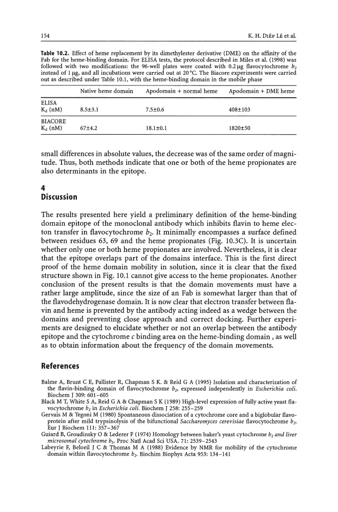
Table 10.2. Effect of heme replacement by its dimethylester derivative (DME) on the affinity of the
Fab for the heme-binding domain. For ELISA tests, the protocol described in Miles et al. (1998) was
followed with two modifications: the 96-well plates were coated with 0.2 ~g flavocytochrome b2
instead of 1 ~g, and all incubations were carried out at 20°C. The Biacore experiments were carried
out as described under Table 10.1, with the heme-binding domain in the mobile phase
Native heme domain
Apodomain + normal heme
Apodomain + DME heme
ELISA
Kd (nM)
8.5±3.1
7.5±0.6
408±103
BIACORE
Kd (nM)
67±4.2
18.1±0.1
1820±50
small differences in absolute values, the decrease was of the same order of magnitude. Thus, both methods indicate that one or both of the heme propionates are
also determinants in the epitope.
4
Discussion
The results presented here yield a preliminary definition of the heme-binding
domain epitope of the monoclonal antibody which inhibits flavin to heme electon transfer in flavocytochrome b2• It minimally encompasses a surface defined
between residues 63, 69 and the heme propionates (Fig. 1O.3C). It is uncertain
whether only one or both heme propionates are involved. Nevertheless, it is clear
that the epitope overlaps part of the domains interface. This is the first direct
proof of the heme domain mobility in solution, since it is clear that the fixed
structure shown in Fig. 10.1 cannot give access to the heme propionates. Another
conclusion of the present results is that the domain movements must have a
rather large amplitude, since the size of an Fab is somewhat larger than that of
the flavodehydrogenase domain. It is now clear that electron transfer between flavin and heme is prevented by the antibody acting indeed as a wedge between the
domains and preventing close approach and correct docking. Further experiments are designed to elucidate whether or not an overlap between the antibody
epitope and the cytochrome c binding area on the heme-binding domain, as well
as to obtain information about the frequency of the domain movements.
References
Balme A, Brunt C E, Pallister R, Chapman S K. & Reid G A (1995) Isolation and characterization of
the flavin-binding domain of flavocytochrome b" expressed independently in Escherichia coli.
Biochem J 309: 601-605
Black M T, White S A, Reid G A & Chapman S K (1989) High-level expression of fully active yeast flavocytochrome b2 in Escherichia coli. Biochem J 258: 255-259
Gervais M & Tegoni M (1980) Spontaneous dissociation of a cytochrome core and a biglobular flavoprotein after mild trypsinolysis of the bifunctional Saccharomyces cerevisiae flavocytochrome b2•
Eur J Biochem 111: 357-367
Guiard B, Groudinsky 0 & Lederer F (1974) Homology between baker's yeast cytochrome b2 and liver
microsomal cytochrome bs. Proc Natl Acad Sci USA. 71: 2539-2543
Labeyrie F, Beloeil J C & Thomas M A (1988) Evidence by NMR for mobility of the cytochrome
domain within flavocytochrome b2• Biochim Biophys Acta 953: 134-141
K. H. DIEP LE et al.
Table 10.2. Effect of heme replacement by its dimethylester derivative (DME) on the affinity of the
Fab for the heme-binding domain. For ELISA tests, the protocol described in Miles et al. (1998) was
followed with two modifications: the 96-well plates were coated with 0.2 ~g flavocytochrome b2
instead of 1 ~g, and all incubations were carried out at 20°C. The Biacore experiments were carried
out as described under Table 10.1, with the heme-binding domain in the mobile phase
Native heme domain
Apodomain + normal heme
Apodomain + DME heme
ELISA
Kd (nM)
8.5±3.1
7.5±0.6
408±103
BIACORE
Kd (nM)
67±4.2
18.1±0.1
1820±50
small differences in absolute values, the decrease was of the same order of magnitude. Thus, both methods indicate that one or both of the heme propionates are
also determinants in the epitope.
4
Discussion
The results presented here yield a preliminary definition of the heme-binding
domain epitope of the monoclonal antibody which inhibits flavin to heme electon transfer in flavocytochrome b2• It minimally encompasses a surface defined
between residues 63, 69 and the heme propionates (Fig. 1O.3C). It is uncertain
whether only one or both heme propionates are involved. Nevertheless, it is clear
that the epitope overlaps part of the domains interface. This is the first direct
proof of the heme domain mobility in solution, since it is clear that the fixed
structure shown in Fig. 10.1 cannot give access to the heme propionates. Another
conclusion of the present results is that the domain movements must have a
rather large amplitude, since the size of an Fab is somewhat larger than that of
the flavodehydrogenase domain. It is now clear that electron transfer between flavin and heme is prevented by the antibody acting indeed as a wedge between the
domains and preventing close approach and correct docking. Further experiments are designed to elucidate whether or not an overlap between the antibody
epitope and the cytochrome c binding area on the heme-binding domain, as well
as to obtain information about the frequency of the domain movements.
References
Balme A, Brunt C E, Pallister R, Chapman S K. & Reid G A (1995) Isolation and characterization of
the flavin-binding domain of flavocytochrome b" expressed independently in Escherichia coli.
Biochem J 309: 601-605
Black M T, White S A, Reid G A & Chapman S K (1989) High-level expression of fully active yeast flavocytochrome b2 in Escherichia coli. Biochem J 258: 255-259
Gervais M & Tegoni M (1980) Spontaneous dissociation of a cytochrome core and a biglobular flavoprotein after mild trypsinolysis of the bifunctional Saccharomyces cerevisiae flavocytochrome b2•
Eur J Biochem 111: 357-367
Guiard B, Groudinsky 0 & Lederer F (1974) Homology between baker's yeast cytochrome b2 and liver
microsomal cytochrome bs. Proc Natl Acad Sci USA. 71: 2539-2543
Labeyrie F, Beloeil J C & Thomas M A (1988) Evidence by NMR for mobility of the cytochrome
domain within flavocytochrome b2• Biochim Biophys Acta 953: 134-141