

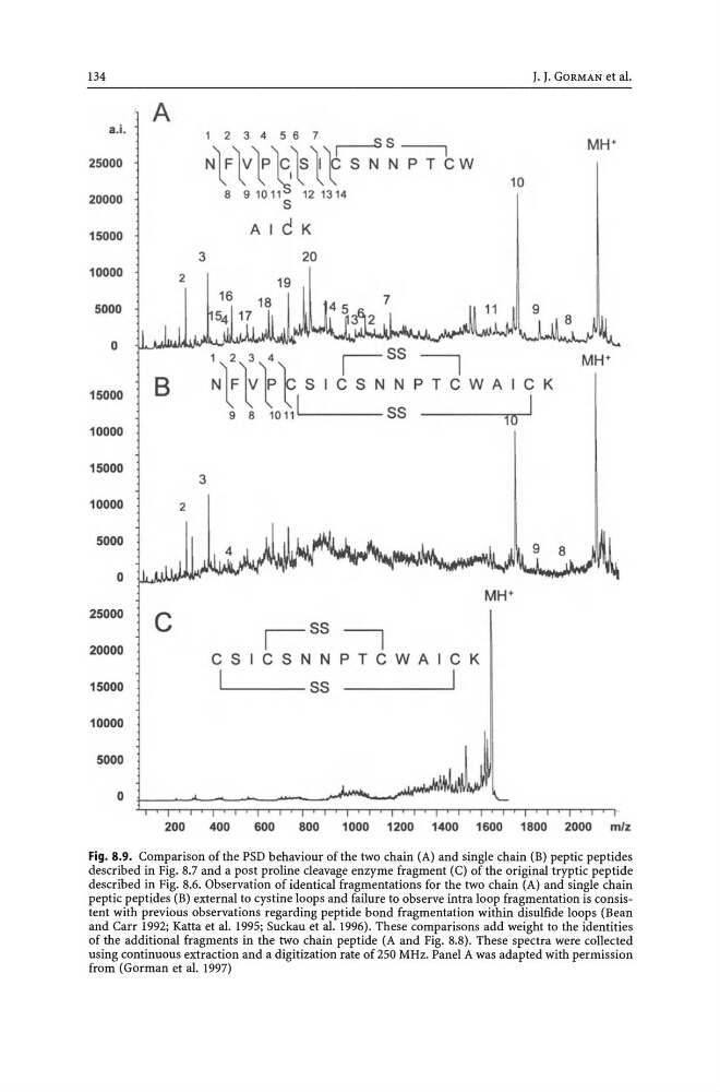
Fig. 8.9. Comparison of the PSD behaviour of the two chain (A) and single chain (B) peptic peptides
described in Fig. 8.7 and a post proline cleavage enzyme fragment (C) of the original tryptic peptide
described in Fig. 8.6. Observation of identical fragmentations for the two chain (A) and single chain
peptic pep tides (B) external to cystine loops and failure to observe intra loop fragmentation is consistent with previous observations regarding peptide bond fragmentation within disulfide loops (Bean
and Carr 1992; Katta et al. 1995; Suckau et al. 1996). These comparisons add weight to the identities
of the additional fragments in the two chain peptide (A and Fig. 8.8). These spectra were collected
using continuous extraction and a digitization rate of 250 MHz. Panel A was adapted with permission
from (Gorman et al. 1997)
described in Fig. 8.7 and a post proline cleavage enzyme fragment (C) of the original tryptic peptide
described in Fig. 8.6. Observation of identical fragmentations for the two chain (A) and single chain
peptic pep tides (B) external to cystine loops and failure to observe intra loop fragmentation is consistent with previous observations regarding peptide bond fragmentation within disulfide loops (Bean
and Carr 1992; Katta et al. 1995; Suckau et al. 1996). These comparisons add weight to the identities
of the additional fragments in the two chain peptide (A and Fig. 8.8). These spectra were collected
using continuous extraction and a digitization rate of 250 MHz. Panel A was adapted with permission
from (Gorman et al. 1997)