

Protein Structure and Dynamics by NMR in Solution
107
A
B
"
c
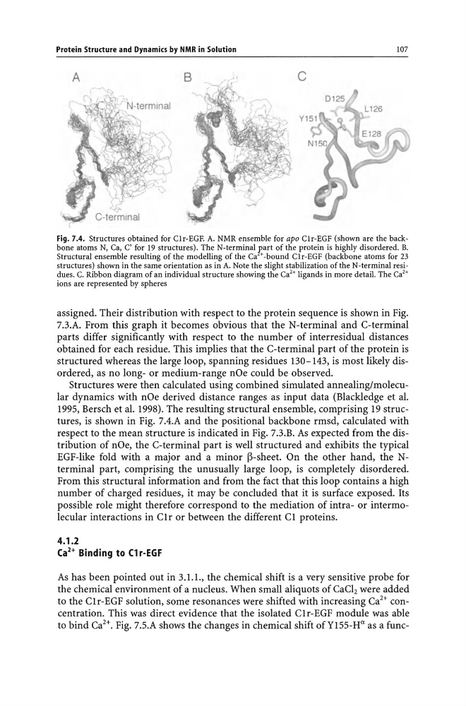
Fig. 7.4. Structures obtained for Clr-EGF. A. NMR ensemble for apo Clr-EGF (shown are the backbone atoms N, Ca, C' for 19 structures). The N-terminal Eart of the protein is highly disordered. B.
Structural ensemble resulting of the modelling of the Ca + -bound Clr-EGF (backbone atoms for 23
structures) shown in the same orientation as in A. Note the slight stabilization of the N-terminal residues. C. Ribbon diagram of an individual structure showing the Ca 2 + ligands in more detail. The Ca'+
ions are represented by spheres
assigned. Their distribution with respect to the protein sequence is shown in Fig.
7.3.A. From this graph it becomes obvious that the N-terminal and C-terminal
parts differ significantly with respect to the number of interresidual distances
obtained for each residue. This implies that the C-terminal part of the protein is
structured whereas the large loop, spanning residues 130-143, is most likely disordered, as no long- or medium-range nOe could be observed.
Structures were then calculated using combined simulated annealing/molecular dynamics with nOe derived distance ranges as input data (Blackledge et al.
1995, Bersch et al. 1998). The resulting structural ensemble, comprising 19 structures, is shown in Fig. 7.4.A and the positional backbone rmsd, calculated with
respect to the mean structure is indicated in Fig. 7.3.B. As expected from the distribution of nOe, the C-terminal part is well structured and exhibits the typical
EGF-like fold with a major and a minor ~-sheet. On the other hand, the Nterminal part, comprising the unusually large loop, is completely disordered.
From this structural information and from the fact that this loop contains a high
number of charged residues, it may be concluded that it is surface exposed. Its
possible role might therefore correspond to the mediation of intra- or intermolecular interactions in C1r or between the different C1 proteins.
4.1.2
(a 2 + Binding to (1 r-EGF
As has been pointed out in 3.1.1., the chemical shift is a very sensitive probe for
the chemical environment of a nucleus. When small aliquots of CaCl2 were added
to the C1r-EGF solution, some resonances were shifted with increasing Ca 2 + concentration. This was direct evidence that the isolated C1r-EGF module was able
to bind Ca 2 +. Fig. 7.5.A shows the changes in chemical shift ofY155-H u as a func-
107
A
B
"
c
Fig. 7.4. Structures obtained for Clr-EGF. A. NMR ensemble for apo Clr-EGF (shown are the backbone atoms N, Ca, C' for 19 structures). The N-terminal Eart of the protein is highly disordered. B.
Structural ensemble resulting of the modelling of the Ca + -bound Clr-EGF (backbone atoms for 23
structures) shown in the same orientation as in A. Note the slight stabilization of the N-terminal residues. C. Ribbon diagram of an individual structure showing the Ca 2 + ligands in more detail. The Ca'+
ions are represented by spheres
assigned. Their distribution with respect to the protein sequence is shown in Fig.
7.3.A. From this graph it becomes obvious that the N-terminal and C-terminal
parts differ significantly with respect to the number of interresidual distances
obtained for each residue. This implies that the C-terminal part of the protein is
structured whereas the large loop, spanning residues 130-143, is most likely disordered, as no long- or medium-range nOe could be observed.
Structures were then calculated using combined simulated annealing/molecular dynamics with nOe derived distance ranges as input data (Blackledge et al.
1995, Bersch et al. 1998). The resulting structural ensemble, comprising 19 structures, is shown in Fig. 7.4.A and the positional backbone rmsd, calculated with
respect to the mean structure is indicated in Fig. 7.3.B. As expected from the distribution of nOe, the C-terminal part is well structured and exhibits the typical
EGF-like fold with a major and a minor ~-sheet. On the other hand, the Nterminal part, comprising the unusually large loop, is completely disordered.
From this structural information and from the fact that this loop contains a high
number of charged residues, it may be concluded that it is surface exposed. Its
possible role might therefore correspond to the mediation of intra- or intermolecular interactions in C1r or between the different C1 proteins.
4.1.2
(a 2 + Binding to (1 r-EGF
As has been pointed out in 3.1.1., the chemical shift is a very sensitive probe for
the chemical environment of a nucleus. When small aliquots of CaCl2 were added
to the C1r-EGF solution, some resonances were shifted with increasing Ca 2 + concentration. This was direct evidence that the isolated C1r-EGF module was able
to bind Ca 2 +. Fig. 7.5.A shows the changes in chemical shift ofY155-H u as a func-