

108
E a.
.e:
Q)
<.>
co
Ql
Q;
:c
'6
~
:c
'"
Oi
C)
·E
Ql
.s;;;;
C)
0.4
0.3
0.2
0.1
0
·0.1
·0.2
·0.3
·0.4
o 40 80 120 160
free Ca2+ I mM
130
140
A
• •
1
-
150
160
residue number
B. BERSCH et al.
170
B
- -1 0.4
03
02
·0.2
·0.3
-0.4
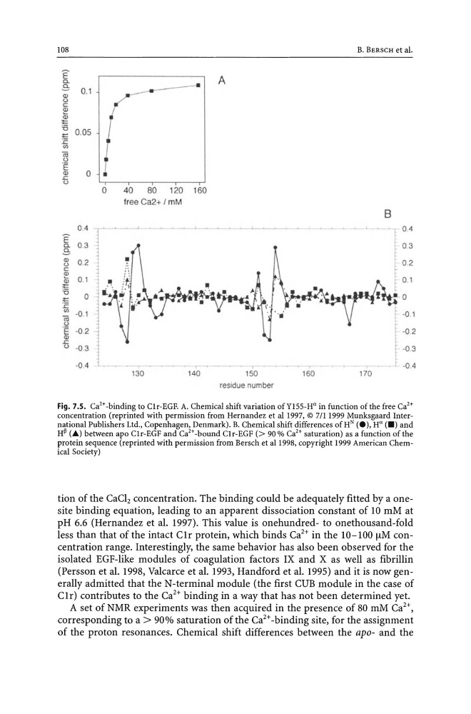
Fig. 7.5. Ca2+·binding to C1r-EGF. A. Chemical shift variation ofYl55·H u in function of the free Ca'+
concentration (reprinted with permission from Hernandez et a! 1997, © 7111999 Munksgaard International Publishers Ltd., Copenhagen, Denmark). B. Chemica! shift differences ofH N (e), H U ( _ ) and
H~ (A) between apo C1r-EGF and Ca'+-bound C1r·EGF (> 90 % Ca'+ saturation) as a function of the
protein sequence (reprinted with permission from Bersch et a11998, copyright 1999 American Chemica! Society)
tion of the CaCh concentration. The binding could be adequately fitted by a onesite binding equation, leading to an apparent dissociation constant of 10 mM at
pH 6.6 (Hernandez et al. 1997). This value is onehundred- to onethousand-fold
less than that of the intact C1r protein, which binds Ca 2 + in the 10-100 [lM concentration range. Interestingly, the same behavior has also been observed for the
isolated EGF-like modules of coagulation factors IX and X as well as fibrillin
(Persson et al. 1998, Valcarce et al. 1993, Handford et al. 1995) and it is now generally admitted that the N-terminal module (the first CUB module in the case of
C1r) contributes to the Ca 2 + binding in a way that has not been determined yet.
A set of NMR experiments was then acquired in the presence of 80 mM Ca 2 +,
corresponding to a > 90% saturation of the Ca 2 + -binding site, for the assignment
of the proton resonances. Chemical shift differences between the apo- and the
E a.
.e:
Q)
<.>
co
Ql
Q;
:c
'6
~
:c
'"
Oi
C)
·E
Ql
.s;;;;
C)
0.4
0.3
0.2
0.1
0
·0.1
·0.2
·0.3
·0.4
o 40 80 120 160
free Ca2+ I mM
130
140
A
• •
1
-
150
160
residue number
B. BERSCH et al.
170
B
- -1 0.4
03
02
·0.2
·0.3
-0.4
Fig. 7.5. Ca2+·binding to C1r-EGF. A. Chemical shift variation ofYl55·H u in function of the free Ca'+
concentration (reprinted with permission from Hernandez et a! 1997, © 7111999 Munksgaard International Publishers Ltd., Copenhagen, Denmark). B. Chemica! shift differences ofH N (e), H U ( _ ) and
H~ (A) between apo C1r-EGF and Ca'+-bound C1r·EGF (> 90 % Ca'+ saturation) as a function of the
protein sequence (reprinted with permission from Bersch et a11998, copyright 1999 American Chemica! Society)
tion of the CaCh concentration. The binding could be adequately fitted by a onesite binding equation, leading to an apparent dissociation constant of 10 mM at
pH 6.6 (Hernandez et al. 1997). This value is onehundred- to onethousand-fold
less than that of the intact C1r protein, which binds Ca 2 + in the 10-100 [lM concentration range. Interestingly, the same behavior has also been observed for the
isolated EGF-like modules of coagulation factors IX and X as well as fibrillin
(Persson et al. 1998, Valcarce et al. 1993, Handford et al. 1995) and it is now generally admitted that the N-terminal module (the first CUB module in the case of
C1r) contributes to the Ca 2 + binding in a way that has not been determined yet.
A set of NMR experiments was then acquired in the presence of 80 mM Ca 2 +,
corresponding to a > 90% saturation of the Ca 2 + -binding site, for the assignment
of the proton resonances. Chemical shift differences between the apo- and the