

106
B. BERSCH et al.
D
0
3.5
0
~
.., .. - .~ · .. f ~ . .. , .:;.
" 0
:. . . . e 4.0
... ~ -
G Arg160
0
•
~ .. - .
•
. : - ..
-..# .
.q <4
4.5
G Ser171
0
•
-
~ ...
-.,
.-, ..
E
~i& ~ . . ! - -
a.
0
0
S
.
5.0
-~ •
N
0 ••
LL
•
5.5
....
.
-
6.0
11 .0
10.5
10.0
9.5
9.0
8.5
8 .0
7 .5
F1 (ppm)
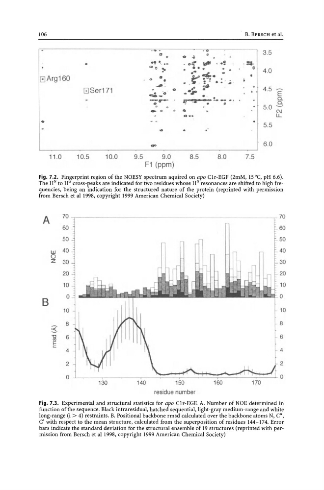
Fig. 7.2. Fingerprint region of the NOESY spectrum aquired on apo Clr-EGF (2mM, 15°C, pH 6.6).
The HN to H a cross-peaks are indicated for two residues whose HN resonances are shifted to high frequencies, being an indication for the structured nature of the protein (reprinted with permission
from Bersch et al1998, copyright 1999 American Chemical Society)
A
70
60
50
w 40
0
z 30
20
10
8
0
10
~
8
'0
6
VJ
E 4
2
0
~-----130
140
150
residue number
170
70
60
50
40
30
20
10
0
10
· 8
6
l:
0
Fig. 7.3. Experimental and structural statistics for apo Clr-EGF. A. Number of NOE determined in
function of the sequence. Black intraresidual, hatched sequential, light-gray medium-range and white
long-range (i > 4) restraints. B. Positional backbone rmsd calculated over the backbone atoms N, C a ,
C' with respect to the mean structure, calculated from the superposition of residues 144-174. Error
bars indicate the standard deviation for the structural ensemble of 19 structures (reprinted with permission from Bersch et al 1998, copyright 1999 American Chemical Society)
B. BERSCH et al.
D
0
3.5
0
~
.., .. - .~ · .. f ~ . .. , .:;.
" 0
:. . . . e 4.0
... ~ -
G Arg160
0
•
~ .. - .
•
. : - ..
-..# .
.q <4
4.5
G Ser171
0
•
-
~ ...
-.,
.-, ..
E
~i& ~ . . ! - -
a.
0
0
S
.
5.0
-~ •
N
0 ••
LL
•
5.5
....
.
-
6.0
11 .0
10.5
10.0
9.5
9.0
8.5
8 .0
7 .5
F1 (ppm)
Fig. 7.2. Fingerprint region of the NOESY spectrum aquired on apo Clr-EGF (2mM, 15°C, pH 6.6).
The HN to H a cross-peaks are indicated for two residues whose HN resonances are shifted to high frequencies, being an indication for the structured nature of the protein (reprinted with permission
from Bersch et al1998, copyright 1999 American Chemical Society)
A
70
60
50
w 40
0
z 30
20
10
8
0
10
~
8
'0
6
VJ
E 4
2
0
~-----130
140
150
residue number
170
70
60
50
40
30
20
10
0
10
· 8
6
l:
0
Fig. 7.3. Experimental and structural statistics for apo Clr-EGF. A. Number of NOE determined in
function of the sequence. Black intraresidual, hatched sequential, light-gray medium-range and white
long-range (i > 4) restraints. B. Positional backbone rmsd calculated over the backbone atoms N, C a ,
C' with respect to the mean structure, calculated from the superposition of residues 144-174. Error
bars indicate the standard deviation for the structural ensemble of 19 structures (reprinted with permission from Bersch et al 1998, copyright 1999 American Chemical Society)