

ferrous state (Fe
3+
! Fe
2+ ). The single electron is delivered from NAD(P)H via
another cofactor, which (depending on the enzyme) is a flavin, an iron-sulfur
protein (ferredoxin) or a cytochrome b 5 . Next, molecular oxygen is bound to give
a Cyt P-450 dioxygen complex. Delivery of a second electron and protonation
forms Compound 0. Protonation cleaves the O–OH bond with expulsion of water
and forms the ultimate oxidizing Fe
4+
¼O species called Compound I, which – as a
strong electrophile – attacks the substrate [1193]. Expulsion of the product (SubO)
reforms the Fe
3+ species and closes the catalytic cycle. Put simply, Cyt P-450
resembles an oxidation by a hypervalent transition metal oxidant (nature’s
permanganate).
Despite the fact that the mechanism of Cyt P-450 enzymes has been intensively
investigated over half a century [1194], many mechanistic details are still poorly
understood and it was only recently, that the existence of an Fe
5+ species was ruled
out [1195].
Aside from the productive cycle, Compound 0 can be formed directly by H 2 O 2
through the so-called ‘peroxide-shunt’. This obviates the necessity for additional
electron-transport components described above, because no single-electron transfer
occurs. However, so far, the use of P-450 enzymes in the peroxygenase-mode is
impeded by limited enzyme stabilities in presence of H 2 O 2 [1196]. Under certain
conditions, Compound 0 may liberate H 2 O 2 , or Compound I may decompose
forming H 2 O, which wastes NAD(P)H in futile cycles, processes which are called
‘uncoupling’. Hence, it is not surprising, that P-450 enzymes are comparatively
slow catalysts with typical TOFs of ~1 s
À1 .
Cyt P-450 enzymes got their name from their hemoprotein character: P stands
for ‘pigment’ and 450 reflects the absorption of the CO-complex at 450 nm. To
date, more than 200,000 distinct Cyt P-450 enzymes are known and these proteins
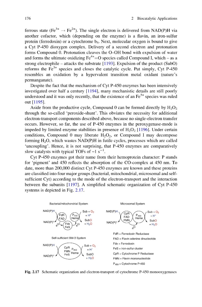
are classified into four major groups (bacterial, mitochondrial, microsomal and selfsufficient Cyt) according to the mode of the electron-transport and the interaction
between the subunits [1197]. A simplified schematic organization of Cyt P-450
systems is depicted in Fig. 2.17.
Sub + O 2
+ H +
NAD(P)H
NAD(P)
+
Fdx
FdR = Ferredoxin Reductase
Fdx = Ferredoxin
P 450 = Cytochrome P-450
CpR
FMN
CpR = Cytochrome P Reductase
CpR
FMN Heme
FAD = Flavin adenine dinucleotide
FMN = Flavin mononucleotide
FeS = iron-sulfur cluster
SubO
+ H 2 O
FdR
Heme
Heme
Bacterial/mitochondrial System
Microsomal System
Self-sufficient BM-3 System
e
e
e
P 450
FeS
FAD
FeS
P 450
P 450
FAD
Sub + O 2
+ H
+
NAD(P)H
NAD(P)
+
SubO
+ H 2 O
Sub + O 2
+ H +
NAD(P)H
NAD(P)
+
SubO
+ H 2 O
Fig. 2.17 Schematic organization and electron-transport of cytochrome P-450 monooxygenases
176
2 Biocatalytic Applications
3+
! Fe
2+ ). The single electron is delivered from NAD(P)H via
another cofactor, which (depending on the enzyme) is a flavin, an iron-sulfur
protein (ferredoxin) or a cytochrome b 5 . Next, molecular oxygen is bound to give
a Cyt P-450 dioxygen complex. Delivery of a second electron and protonation
forms Compound 0. Protonation cleaves the O–OH bond with expulsion of water
and forms the ultimate oxidizing Fe
4+
¼O species called Compound I, which – as a
strong electrophile – attacks the substrate [1193]. Expulsion of the product (SubO)
reforms the Fe
3+ species and closes the catalytic cycle. Put simply, Cyt P-450
resembles an oxidation by a hypervalent transition metal oxidant (nature’s
permanganate).
Despite the fact that the mechanism of Cyt P-450 enzymes has been intensively
investigated over half a century [1194], many mechanistic details are still poorly
understood and it was only recently, that the existence of an Fe
5+ species was ruled
out [1195].
Aside from the productive cycle, Compound 0 can be formed directly by H 2 O 2
through the so-called ‘peroxide-shunt’. This obviates the necessity for additional
electron-transport components described above, because no single-electron transfer
occurs. However, so far, the use of P-450 enzymes in the peroxygenase-mode is
impeded by limited enzyme stabilities in presence of H 2 O 2 [1196]. Under certain
conditions, Compound 0 may liberate H 2 O 2 , or Compound I may decompose
forming H 2 O, which wastes NAD(P)H in futile cycles, processes which are called
‘uncoupling’. Hence, it is not surprising, that P-450 enzymes are comparatively
slow catalysts with typical TOFs of ~1 s
À1 .
Cyt P-450 enzymes got their name from their hemoprotein character: P stands
for ‘pigment’ and 450 reflects the absorption of the CO-complex at 450 nm. To
date, more than 200,000 distinct Cyt P-450 enzymes are known and these proteins
are classified into four major groups (bacterial, mitochondrial, microsomal and selfsufficient Cyt) according to the mode of the electron-transport and the interaction
between the subunits [1197]. A simplified schematic organization of Cyt P-450
systems is depicted in Fig. 2.17.
Sub + O 2
+ H +
NAD(P)H
NAD(P)
+
Fdx
FdR = Ferredoxin Reductase
Fdx = Ferredoxin
P 450 = Cytochrome P-450
CpR
FMN
CpR = Cytochrome P Reductase
CpR
FMN Heme
FAD = Flavin adenine dinucleotide
FMN = Flavin mononucleotide
FeS = iron-sulfur cluster
SubO
+ H 2 O
FdR
Heme
Heme
Bacterial/mitochondrial System
Microsomal System
Self-sufficient BM-3 System
e
e
e
P 450
FeS
FAD
FeS
P 450
P 450
FAD
Sub + O 2
+ H
+
NAD(P)H
NAD(P)
+
SubO
+ H 2 O
Sub + O 2
+ H +
NAD(P)H
NAD(P)
+
SubO
+ H 2 O
Fig. 2.17 Schematic organization and electron-transport of cytochrome P-450 monooxygenases
176
2 Biocatalytic Applications