

6 In Situ Hybridization for DNA: Fluorescent Probe
83
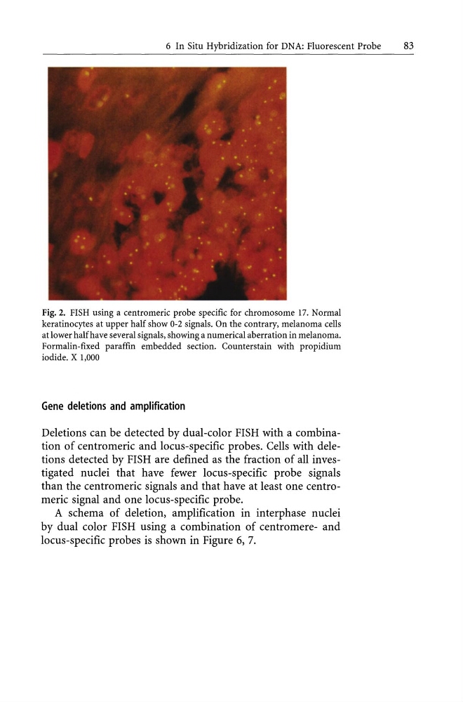
Fig. 2. FISH using a centromeric probe specific for chromosome 17. Normal
keratinocytes at upper half show 0-2 signals. On the contrary, melanoma cells
at lower halfhave several signals, showing a numerical aberration in melanoma.
Formalin-fixed paraffin embedded section. Counterstain with propidium
iodide. X 1,000
Gene deletions and amplification
Deletions can be detected by dual-color FISH with a combination of centromeric and locus-specific probes. Cells with deletions detected by FISH are defined as the fraction of all investigated nuclei that have fewer locus-specific probe signals
than the centromeric signals and that have at least one centromeric signal and one locus-specific probe.
A schema of deletion, amplification in interphase nuclei
by dual color FISH using a combination of centromere- and
locus-specific probes is shown in Figure 6, 7.
83
Fig. 2. FISH using a centromeric probe specific for chromosome 17. Normal
keratinocytes at upper half show 0-2 signals. On the contrary, melanoma cells
at lower halfhave several signals, showing a numerical aberration in melanoma.
Formalin-fixed paraffin embedded section. Counterstain with propidium
iodide. X 1,000
Gene deletions and amplification
Deletions can be detected by dual-color FISH with a combination of centromeric and locus-specific probes. Cells with deletions detected by FISH are defined as the fraction of all investigated nuclei that have fewer locus-specific probe signals
than the centromeric signals and that have at least one centromeric signal and one locus-specific probe.
A schema of deletion, amplification in interphase nuclei
by dual color FISH using a combination of centromere- and
locus-specific probes is shown in Figure 6, 7.