

84
MORIMASA MATSUTA AND MAYUMI MATS UTA
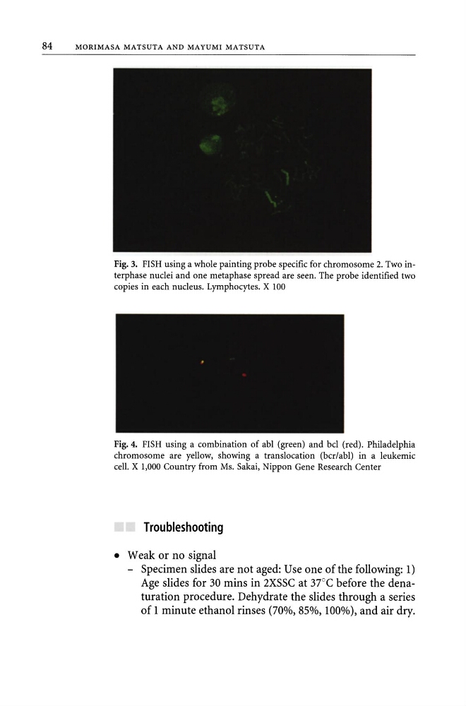
Fig. 3. FISH using a whole painting probe specific for chromosome 2. Two interphase nuclei and one metaphase spread are seen. The probe identified two
copies in each nucleus. Lymphocytes. X 100
Fig. 4. FISH using a combination of abl (green) and bcl (red). Philadelphia
chromosome are yellow, showing a translocation (bcr/abl) in a leukemic
cell. X 1,000 Country from Ms. Sakai, Nippon Gene Research Center
Troubleshooting
• Weak or no signal
- Specimen slides are not aged: Use one of the following: 1)
Age slides for 30 mins in 2XSSC at 37°C before the denaturation procedure. Dehydrate the slides through a series
of 1 minute ethanol rinses (70%, 85%,100%), and air dry.
MORIMASA MATSUTA AND MAYUMI MATS UTA
Fig. 3. FISH using a whole painting probe specific for chromosome 2. Two interphase nuclei and one metaphase spread are seen. The probe identified two
copies in each nucleus. Lymphocytes. X 100
Fig. 4. FISH using a combination of abl (green) and bcl (red). Philadelphia
chromosome are yellow, showing a translocation (bcr/abl) in a leukemic
cell. X 1,000 Country from Ms. Sakai, Nippon Gene Research Center
Troubleshooting
• Weak or no signal
- Specimen slides are not aged: Use one of the following: 1)
Age slides for 30 mins in 2XSSC at 37°C before the denaturation procedure. Dehydrate the slides through a series
of 1 minute ethanol rinses (70%, 85%,100%), and air dry.