

82
MORIMASA MATSUTA AND MAYUMI MATS UTA
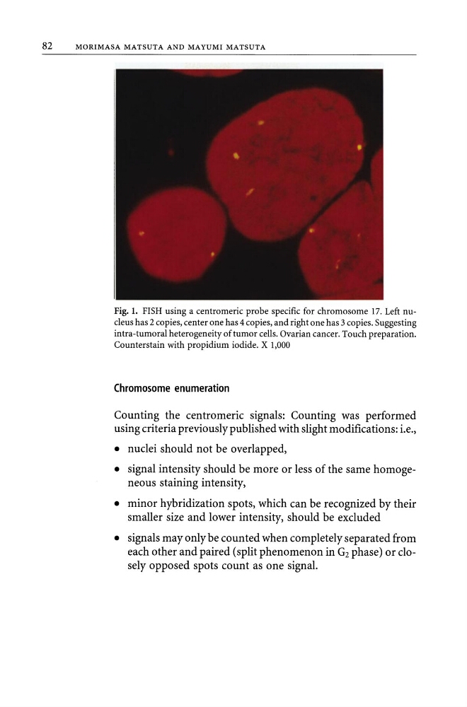
Fig. 1. FISH using a centromeric probe specific for chromosome 17. Left nucleus has 2 copies, center one has 4 copies, and right one has 3 copies. Suggesting
intra-tumoral heterogeneity of tumor cells. Ovarian cancer. Touch preparation.
Counterstain with propidium iodide. X 1,000
Chromosome enumeration
Counting the centromeric signals: Counting was performed
using criteria previously published with slight modifications: i.e.,
• nuclei should not be overlapped,
• signal intensity should be more or less of the same homogeneous staining intensity,
• minor hybridization spots, which can be recognized by their
smaller size and lower intensity, should be excluded
• signals may only be counted when completely separated from
each other and paired (split phenomenon in Gz phase) or closely opposed spots count as one signal.
MORIMASA MATSUTA AND MAYUMI MATS UTA
Fig. 1. FISH using a centromeric probe specific for chromosome 17. Left nucleus has 2 copies, center one has 4 copies, and right one has 3 copies. Suggesting
intra-tumoral heterogeneity of tumor cells. Ovarian cancer. Touch preparation.
Counterstain with propidium iodide. X 1,000
Chromosome enumeration
Counting the centromeric signals: Counting was performed
using criteria previously published with slight modifications: i.e.,
• nuclei should not be overlapped,
• signal intensity should be more or less of the same homogeneous staining intensity,
• minor hybridization spots, which can be recognized by their
smaller size and lower intensity, should be excluded
• signals may only be counted when completely separated from
each other and paired (split phenomenon in Gz phase) or closely opposed spots count as one signal.