

4 In Situ Nick End-Labeling: Electron Microscopical
39
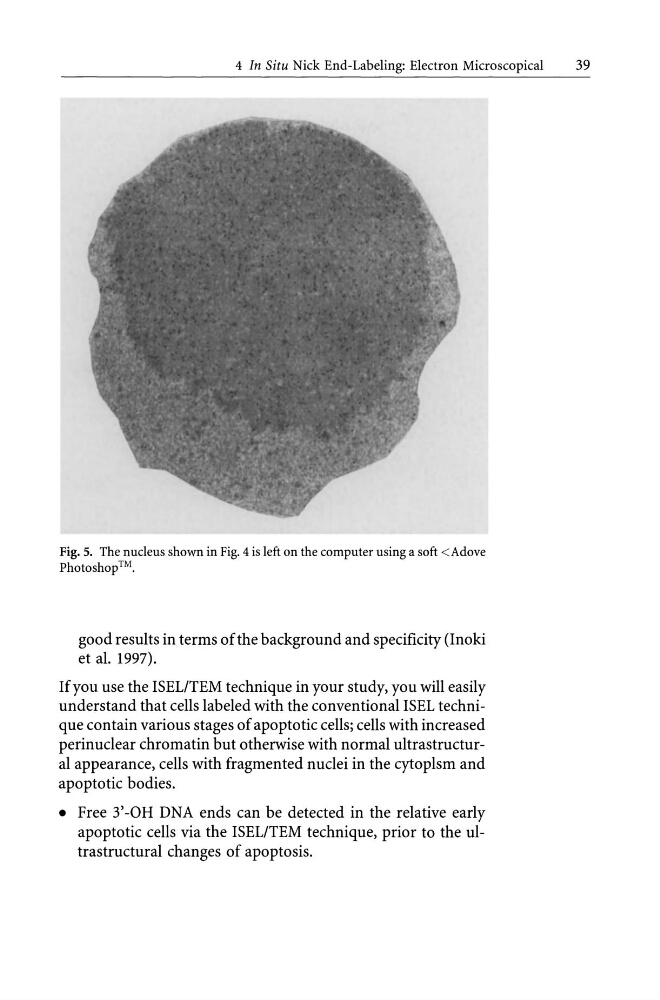
Fig. 5. The nucleus shown in Fig. 4 is left on the computer using a soft
good results in terms of the background and specificity (Inoki
et al. 1997).
If you use the ISELITEM technique in your study, you will easily
understand that cells labeled with the conventional ISEL technique contain various stages of apoptotic cells; cells with increased
perinuclear chromatin but otherwise with normal ultrastructural appearance, cells with fragmented nuclei in the cytoplsm and
apoptotic bodies.
• Free 3' -OH DNA ends can be detected in the relative early
apoptotic cells via the ISELITEM technique, prior to the ultrastructural changes of apoptosis.
39
Fig. 5. The nucleus shown in Fig. 4 is left on the computer using a soft
good results in terms of the background and specificity (Inoki
et al. 1997).
If you use the ISELITEM technique in your study, you will easily
understand that cells labeled with the conventional ISEL technique contain various stages of apoptotic cells; cells with increased
perinuclear chromatin but otherwise with normal ultrastructural appearance, cells with fragmented nuclei in the cytoplsm and
apoptotic bodies.
• Free 3' -OH DNA ends can be detected in the relative early
apoptotic cells via the ISELITEM technique, prior to the ultrastructural changes of apoptosis.