

112
TAKASHI SAWAI AND MIWA UZUKI
and biochemistry in the field of pathology and developmental
biology.
Applications
• Developmental biology
Fig. 1 shows the mRNA of type II collagen in the limb of a
chick embryo at stage 28 demonstrated by 3H labeled probe
in situ hybridization. Observation of the development process
clearly shows that the bones grow from both peripheral regions formed in the limbs.
• Inflammation
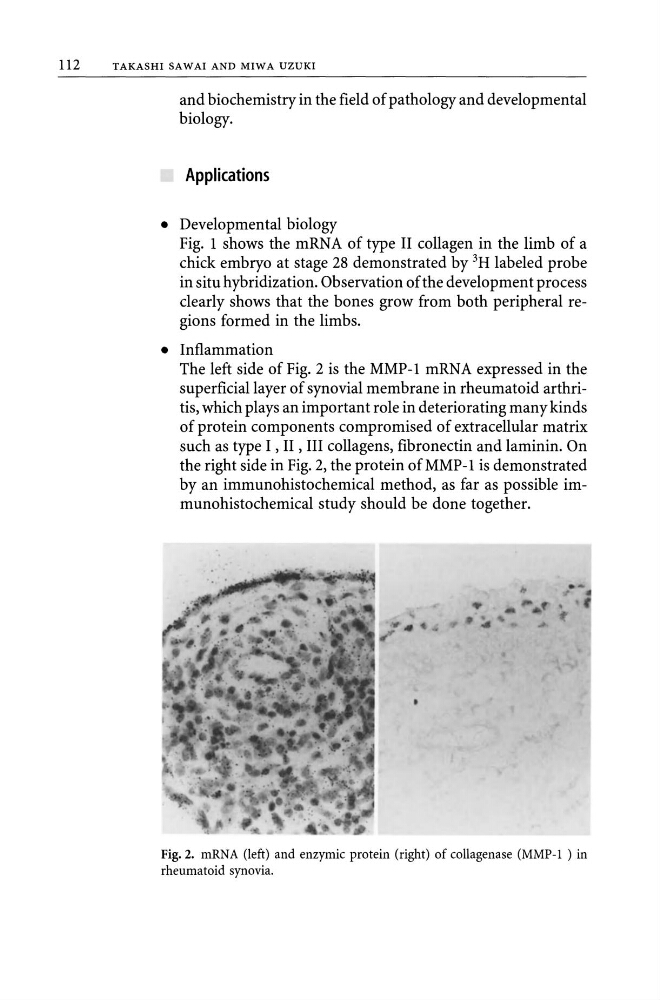
The left side of Fig. 2 is the MMP-l mRNA expressed in the
superficial layer of synovial membrane in rheumatoid arthritis, which plays an important role in deteriorating many kinds
of protein components compromised of extracellular matrix
such as type I , II , III collagens, fibronectin and laminin. On
the right side in Fig. 2, the protein of MMP-l is demonstrated
by an immunohistochemical method, as far as possible immunohistochemical study should be done together.
Fig. 2. mRNA (left) and enzymic protein (right) of collagenase (MMP-l ) in
rheumatoid synovia.
TAKASHI SAWAI AND MIWA UZUKI
and biochemistry in the field of pathology and developmental
biology.
Applications
• Developmental biology
Fig. 1 shows the mRNA of type II collagen in the limb of a
chick embryo at stage 28 demonstrated by 3H labeled probe
in situ hybridization. Observation of the development process
clearly shows that the bones grow from both peripheral regions formed in the limbs.
• Inflammation
The left side of Fig. 2 is the MMP-l mRNA expressed in the
superficial layer of synovial membrane in rheumatoid arthritis, which plays an important role in deteriorating many kinds
of protein components compromised of extracellular matrix
such as type I , II , III collagens, fibronectin and laminin. On
the right side in Fig. 2, the protein of MMP-l is demonstrated
by an immunohistochemical method, as far as possible immunohistochemical study should be done together.
Fig. 2. mRNA (left) and enzymic protein (right) of collagenase (MMP-l ) in
rheumatoid synovia.