

282
Assay of Some Common Hydrobiological Techniques
tives and 15 oculars, using the ocular screen shown in Fig. 6.5, while the native
screen of the microscope might also be applied. With this method, it is very
important to empirically select the proper actinide orange concentration
during staining, as well as the duration of staining and filtration to attain the
optimal degree of coloration in the preparation in which the field of the microscope remains dark, while the bacteria are a shining bright blue.
The cells of planktonic bacteria are enumerated and sized in the same
preparation. For their sizing, it is possible to use enlarged microphotographs
of preparations with a dimensional scale. For good statistics, approximately
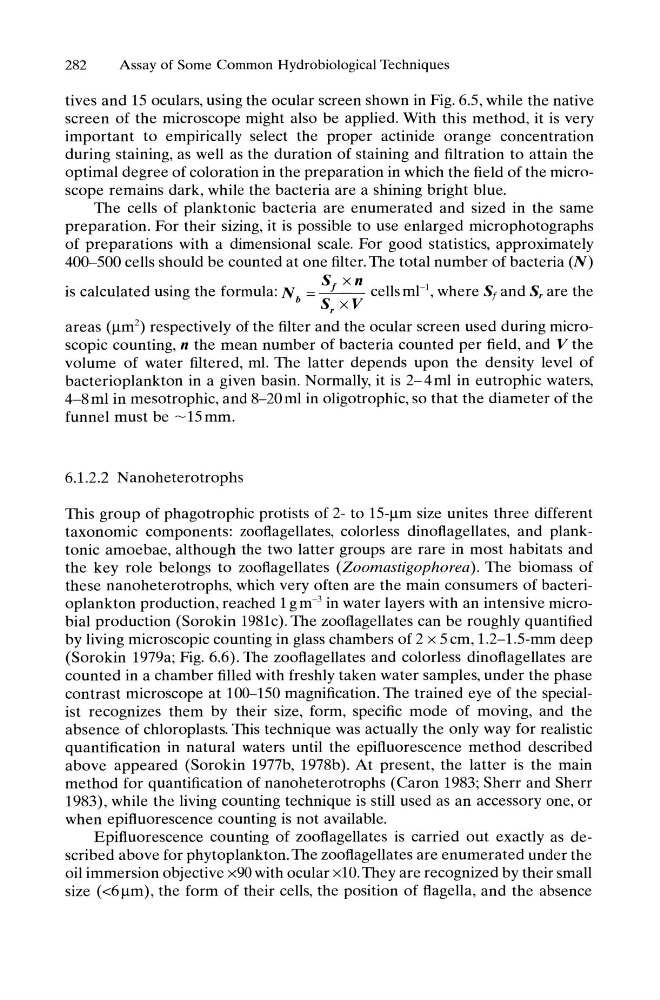
400-500 cells should be counted at one filter. The total number of bacteria (N)
S xn
is calculated using the formula: N b = f
cells ml- 1 , where Sf and Sr are the
S xV
r
areas (~m2) respectively of the filter and the ocular screen used during microscopic counting, n the mean number of bacteria counted per field, and V the
volume of water filtered, ml. The latter depends upon the density level of
bacterioplankton in a given basin. Normally, it is 2-4ml in eutrophic waters,
4-8ml in mesotrophic, and 8-20ml in oligotrophic, so that the diameter of the
funnel must be ~ 15 mm.
6.1.2.2 N anoheterotrophs
This group of phagotrophic protists of 2- to 15-~m size unites three different
taxonomic components: zooflagellates, colorless dinoflagellates, and planktonic amoebae, although the two latter groups are rare in most habitats and
the key role belongs to zooflagellates (Zoomastigophorea). The biomass of
these nanoheterotrophs, which very often are the main consumers of bacterioplankton production, reached 1 gm- 3 in water layers with an intensive microbial production (Sorokin 1981c). The zooflagellates can be roughly quantified
by living microscopic counting in glass chambers of 2 x 5 cm, 1.2-1.5-mm deep
(Sorokin 1979a; Fig. 6.6). The zooflagellates and colorless dinoflagellates are
counted in a chamber filled with freshly taken water samples, under the phase
contrast microscope at 100-150 magnification. The trained eye of the specialist recognizes them by their size, form, specific mode of moving, and the
absence of chloroplasts. This technique was actually the only way for realistic
quantification in natural waters until the epifluorescence method described
above appeared (Sorokin 1977b, 1978b). At present, the latter is the main
method for quantification of nanoheterotrophs (Caron 1983; Sherr and Sherr
1983), while the living counting technique is still used as an accessory one, or
when epifluorescence counting is not available.
Epifluorescence counting of zooflagellates is carried out exactly as described above for phytoplankton. The zooflagellates are enumerated under the
oil immersion objective x90 with ocular xl O. They are recognized by their small
size «6 ~m), the form of their cells, the position of flagella, and the absence
Assay of Some Common Hydrobiological Techniques
tives and 15 oculars, using the ocular screen shown in Fig. 6.5, while the native
screen of the microscope might also be applied. With this method, it is very
important to empirically select the proper actinide orange concentration
during staining, as well as the duration of staining and filtration to attain the
optimal degree of coloration in the preparation in which the field of the microscope remains dark, while the bacteria are a shining bright blue.
The cells of planktonic bacteria are enumerated and sized in the same
preparation. For their sizing, it is possible to use enlarged microphotographs
of preparations with a dimensional scale. For good statistics, approximately
400-500 cells should be counted at one filter. The total number of bacteria (N)
S xn
is calculated using the formula: N b = f
cells ml- 1 , where Sf and Sr are the
S xV
r
areas (~m2) respectively of the filter and the ocular screen used during microscopic counting, n the mean number of bacteria counted per field, and V the
volume of water filtered, ml. The latter depends upon the density level of
bacterioplankton in a given basin. Normally, it is 2-4ml in eutrophic waters,
4-8ml in mesotrophic, and 8-20ml in oligotrophic, so that the diameter of the
funnel must be ~ 15 mm.
6.1.2.2 N anoheterotrophs
This group of phagotrophic protists of 2- to 15-~m size unites three different
taxonomic components: zooflagellates, colorless dinoflagellates, and planktonic amoebae, although the two latter groups are rare in most habitats and
the key role belongs to zooflagellates (Zoomastigophorea). The biomass of
these nanoheterotrophs, which very often are the main consumers of bacterioplankton production, reached 1 gm- 3 in water layers with an intensive microbial production (Sorokin 1981c). The zooflagellates can be roughly quantified
by living microscopic counting in glass chambers of 2 x 5 cm, 1.2-1.5-mm deep
(Sorokin 1979a; Fig. 6.6). The zooflagellates and colorless dinoflagellates are
counted in a chamber filled with freshly taken water samples, under the phase
contrast microscope at 100-150 magnification. The trained eye of the specialist recognizes them by their size, form, specific mode of moving, and the
absence of chloroplasts. This technique was actually the only way for realistic
quantification in natural waters until the epifluorescence method described
above appeared (Sorokin 1977b, 1978b). At present, the latter is the main
method for quantification of nanoheterotrophs (Caron 1983; Sherr and Sherr
1983), while the living counting technique is still used as an accessory one, or
when epifluorescence counting is not available.
Epifluorescence counting of zooflagellates is carried out exactly as described above for phytoplankton. The zooflagellates are enumerated under the
oil immersion objective x90 with ocular xl O. They are recognized by their small
size «6 ~m), the form of their cells, the position of flagella, and the absence