

S. Pantoja . S. Wakeham
a
H
0
I I I
b
I ~ 1
H 2 N - C - C -
H
0
I I I
NH - C - C - O H
I
R
R
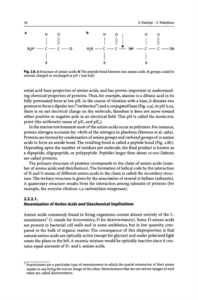
Fig. 2.S. a Structure of amino acids; b The peptide bond between two amino acids. R-groups could be
neutral, charged or uncharged at pH 7 (see text)
ential acid-base properties of amino acids, and has proven important in understanding chemical properties of proteins. Thus, for example, alanine is a dibasic acid in its
fully protonated form at low pH. In the course of titration with a base, it donates two
protons to form a dipolar ion ("zwitlerion") and a conjugated base (Fig. 2.9). At pH 6.02,
there is no net electrical charge on the molecule, therefore it does not move toward
either positive or negative pole in an electrical field. This pH is called the isoelectric
point (the arithmetic mean of pKI and pK 2 ).
In the marine environment most of the amino acids occur as polymers. For instance,
protein nitrogen accounts for >80% of the nitrogen in plankton (Parsons et al. 1984).
Proteins are formed by condensation of amino groups and carboxyl groups of a-amino
acids to form an amide bond. The resulting bond is called a peptide bond (Fig. 2.8b).
Depending upon the number of residues per molecule, the final product is known as
a dipeptide, oligopeptide, or polypeptide. Peptides larger than about 10 000 Daltons
are called proteins.
The primary structure of proteins corresponds to the chain of amino acids (number of amino acids and distribution). The formation of helical coils by the interaction
of Hand 0 atoms of different amino acids in the chain is called the secondary structure. The tertiary structure is given by the association of several a-helixes (subunits).
A quaternary structure results from the interaction among subunits of proteins (for
example, the enzyme ribulose-l,s-carboxylase-oxygenase).
2.2.2.1.
Racemization of Amino Acids and Geochemical Implications
Amino acids commonly found in living organisms consist almost entirely of the Lenantiomers2 (L stands for levorotatory, D for dextrorotatory). Some D-amino acids
are present in bacterial cell walls and in some antibiotics, but in low quantity compared to the bulk of organic matter. The consequence of this disproportion is that
natural amino acids are optically active (except for glycine) and under polarized light
rotate the plane to the left. A racemic mixture would be optically inactive since it contains equal amounts of D- and L-amino acids.
2 Enantiomers are a particular type of stereoisomers in which the spatial orientation of their atoms
results in one being the mirror image of the other. Stereoisomers that are not mirror images of each
other are called diastereomers.
a
H
0
I I I
b
I ~ 1
H 2 N - C - C -
H
0
I I I
NH - C - C - O H
I
R
R
Fig. 2.S. a Structure of amino acids; b The peptide bond between two amino acids. R-groups could be
neutral, charged or uncharged at pH 7 (see text)
ential acid-base properties of amino acids, and has proven important in understanding chemical properties of proteins. Thus, for example, alanine is a dibasic acid in its
fully protonated form at low pH. In the course of titration with a base, it donates two
protons to form a dipolar ion ("zwitlerion") and a conjugated base (Fig. 2.9). At pH 6.02,
there is no net electrical charge on the molecule, therefore it does not move toward
either positive or negative pole in an electrical field. This pH is called the isoelectric
point (the arithmetic mean of pKI and pK 2 ).
In the marine environment most of the amino acids occur as polymers. For instance,
protein nitrogen accounts for >80% of the nitrogen in plankton (Parsons et al. 1984).
Proteins are formed by condensation of amino groups and carboxyl groups of a-amino
acids to form an amide bond. The resulting bond is called a peptide bond (Fig. 2.8b).
Depending upon the number of residues per molecule, the final product is known as
a dipeptide, oligopeptide, or polypeptide. Peptides larger than about 10 000 Daltons
are called proteins.
The primary structure of proteins corresponds to the chain of amino acids (number of amino acids and distribution). The formation of helical coils by the interaction
of Hand 0 atoms of different amino acids in the chain is called the secondary structure. The tertiary structure is given by the association of several a-helixes (subunits).
A quaternary structure results from the interaction among subunits of proteins (for
example, the enzyme ribulose-l,s-carboxylase-oxygenase).
2.2.2.1.
Racemization of Amino Acids and Geochemical Implications
Amino acids commonly found in living organisms consist almost entirely of the Lenantiomers2 (L stands for levorotatory, D for dextrorotatory). Some D-amino acids
are present in bacterial cell walls and in some antibiotics, but in low quantity compared to the bulk of organic matter. The consequence of this disproportion is that
natural amino acids are optically active (except for glycine) and under polarized light
rotate the plane to the left. A racemic mixture would be optically inactive since it contains equal amounts of D- and L-amino acids.
2 Enantiomers are a particular type of stereoisomers in which the spatial orientation of their atoms
results in one being the mirror image of the other. Stereoisomers that are not mirror images of each
other are called diastereomers.