

R. Cecchi· G. Ghermandi
effects are to use low Z and thin targets (of the order of 1 mg cm- 2 , in which the energy loss of proton beam and the X-ray attenuation are negligible) which are suitable
for limiting SEB and BP emissions (Ghermandi et al.I996).
The most widely used accelerators for PIXE experiments are single-ended Van de
Graaff machines. The PIXE set-up used for environmental samples at the National
Institute of Nuclear Physics (INFN) Laboratory of Legnaro (Padova, Italy) is here described (Aprilesi et al. 1984). The incident particles are protons accelerated to 1.8 MeV.
The beam coming from the Van de Graaff is stabilized passing through a slit system
into a deflector magnet, then driven in the line under high vacuum (10 -6 Torr) by electrostatic equipments and lenses, up to the irradiation chamber. To obtain a homogeneous circular beam spot at the target, the beam is diffused through a Ni foil,
450 f..lm cm- 2 , then carbon collimators pick off its central part (approximately 1.5 cm
diameter). The Si(Li) detector (resolution 150 eV at 5.9 keV, solid angle between the
target and the sensitive detector area 0.0198 sr) is placed at 135
0
from the beam and in
front of the target. These measurement conditions limit the X-ray attenuation in the
target. A Faraday cup is placed beyond the sample, at the end of the line, in order to
measure the charge collected on the target. The resulting PIXE spectrum is collected
in a multi channel analyser and then examined by suitable computer codes, in order
to obtain the element concentration in the exposed target.
Mylar absorbers of various thickness or a funny filter (absorber with a central hole)
may be placed in front of the detector. The mylar absorbers reduce the intensity of the
emissions of exceptionally abundant light elements, as it may occur in sea and freshwater samples. Funny filters are preferable for aerosol specimens, because they balance the intensity between the low and high energy regions of the spectrum.
Pore water and mainly sediment samples, in which some medium-heavy elements
are in general particularly abundant, may require the use of absorbers that selectively
attenuate the most intense lines.
19.3
Sensitivity and Detection Limits of the Technique
PIXE intrinsic sensitivity is about 1 ppm of the exposed sample that, in a suitably thin
target (for example 100 f..lg cm- 2 of carbon), corresponds to very small amount (picograms) of an element. The sensitivity of the PIXE measure for a given atomic number
depends on detector parameters, target composition, ion beam and measurement conditions (Cahill 1975). All these factors may be optimized to improve the technique's
sensitivity. In a given set up and unvariable measurement conditions, the target features (thickness, characteristic ionization cross section of the elements both of the
sample and of its backing) determine the behaviour of the yield versus atomic number. The highest sensitivity is obtained for 20 < Z < 40 and Z > 75 and with low proton energies (1-3 MeV). These highest yield regions correspond to the detection limit
minima. The sensitivity in a PIXE spectrum is determined by the signal/noise ratio of
the detected emission. The detection limit is generally estimated as the number of
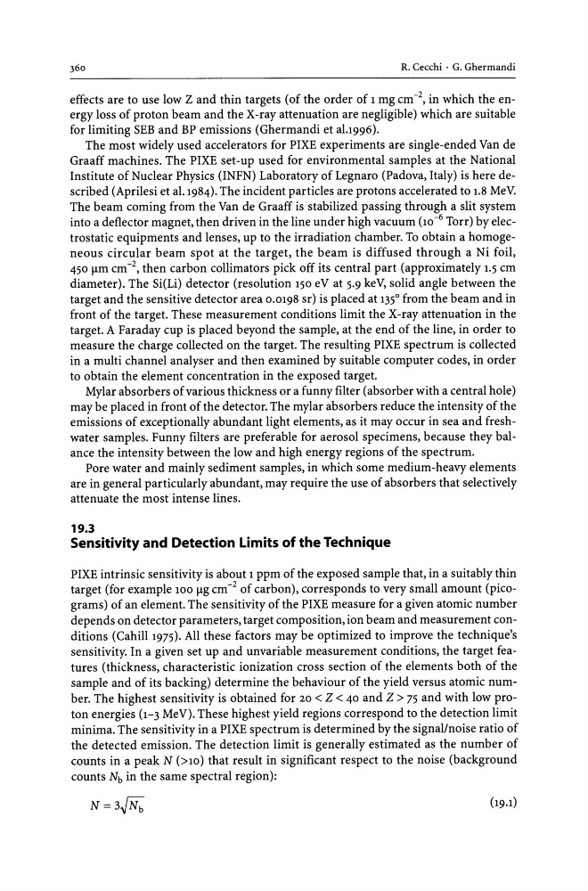
counts in a peak N (>10) that result in significant respect to the noise (background
counts Nb in the same spectral region):
N=3~
effects are to use low Z and thin targets (of the order of 1 mg cm- 2 , in which the energy loss of proton beam and the X-ray attenuation are negligible) which are suitable
for limiting SEB and BP emissions (Ghermandi et al.I996).
The most widely used accelerators for PIXE experiments are single-ended Van de
Graaff machines. The PIXE set-up used for environmental samples at the National
Institute of Nuclear Physics (INFN) Laboratory of Legnaro (Padova, Italy) is here described (Aprilesi et al. 1984). The incident particles are protons accelerated to 1.8 MeV.
The beam coming from the Van de Graaff is stabilized passing through a slit system
into a deflector magnet, then driven in the line under high vacuum (10 -6 Torr) by electrostatic equipments and lenses, up to the irradiation chamber. To obtain a homogeneous circular beam spot at the target, the beam is diffused through a Ni foil,
450 f..lm cm- 2 , then carbon collimators pick off its central part (approximately 1.5 cm
diameter). The Si(Li) detector (resolution 150 eV at 5.9 keV, solid angle between the
target and the sensitive detector area 0.0198 sr) is placed at 135
0
from the beam and in
front of the target. These measurement conditions limit the X-ray attenuation in the
target. A Faraday cup is placed beyond the sample, at the end of the line, in order to
measure the charge collected on the target. The resulting PIXE spectrum is collected
in a multi channel analyser and then examined by suitable computer codes, in order
to obtain the element concentration in the exposed target.
Mylar absorbers of various thickness or a funny filter (absorber with a central hole)
may be placed in front of the detector. The mylar absorbers reduce the intensity of the
emissions of exceptionally abundant light elements, as it may occur in sea and freshwater samples. Funny filters are preferable for aerosol specimens, because they balance the intensity between the low and high energy regions of the spectrum.
Pore water and mainly sediment samples, in which some medium-heavy elements
are in general particularly abundant, may require the use of absorbers that selectively
attenuate the most intense lines.
19.3
Sensitivity and Detection Limits of the Technique
PIXE intrinsic sensitivity is about 1 ppm of the exposed sample that, in a suitably thin
target (for example 100 f..lg cm- 2 of carbon), corresponds to very small amount (picograms) of an element. The sensitivity of the PIXE measure for a given atomic number
depends on detector parameters, target composition, ion beam and measurement conditions (Cahill 1975). All these factors may be optimized to improve the technique's
sensitivity. In a given set up and unvariable measurement conditions, the target features (thickness, characteristic ionization cross section of the elements both of the
sample and of its backing) determine the behaviour of the yield versus atomic number. The highest sensitivity is obtained for 20 < Z < 40 and Z > 75 and with low proton energies (1-3 MeV). These highest yield regions correspond to the detection limit
minima. The sensitivity in a PIXE spectrum is determined by the signal/noise ratio of
the detected emission. The detection limit is generally estimated as the number of
counts in a peak N (>10) that result in significant respect to the noise (background
counts Nb in the same spectral region):
N=3~