

368
a
L. Pellerito· R. Barbieri· R. Di Stefano· M. Scopelliti . C. Pellerito· T. Fiore· F. Triolo
p
/1\
b
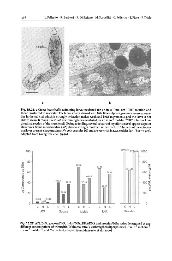
Fig. 15.26. a Ciona intestinalis swimming larva incubated for 1 h in 10- 7 mol dm -3 TBT solution and
then transferred to sea water. The larva, vitally stained with Nile Blue sulphate, presents severe anomalies in the tail (ta) which is strongly twisted; it makes weak and brief movements, and the larva is not
able to swim; b Ciona intestinalis swimming larva incubated for 1 h in 10 -7 mol dm -3 TBT solution. Longitudinal section of the muscle cell. Owing to folding, several sectors of myofibrils (m *j) appear as point
structures. Some mitochondria (m*) show a strongly modified ultrastructure. The cells of the ectodermal layer present a large nucleus (N), yolk granules (G) and are very rich in r.e.r. vesicles (er). (Bar = 1 flm),
adapted from Gianguzza et al. (1996)
100
990 ±45 970 ±50 1 000
« 80
800
Z
70±6
't:
0
67±5
\C
C'I
634
"C
:::L
60
56±6
~
600S
-'tl
ll~
~
.::
48 i5
~.
:::>
IT~
0
40B
37;1;4
Co 40
400 ;B
E
0
0
u
z
C'I
18 i 2
l>
:::L
20
200
3tO.2
2tO.2
ltO.l
0
',' ',' ','
0
C H L
C H L
C H L
C H L
C H L
AlP
Gucose
Lipids
RNA
Proteins
Fig. 15.27. ATP/DNA, glucoselDNA, lipids/DNA, RNA/DNA and proteins/DNA ratios determined at two
different concentrations of tributyltin(IV)[meso-tetra(4-carboxyphenyl)porphinate): H = 10- 5 mol dm-\
L = 10 -7 mol dm - 3; and C = control, adapted from Mansueto et al. (2000)
a
L. Pellerito· R. Barbieri· R. Di Stefano· M. Scopelliti . C. Pellerito· T. Fiore· F. Triolo
p
/1\
b
Fig. 15.26. a Ciona intestinalis swimming larva incubated for 1 h in 10- 7 mol dm -3 TBT solution and
then transferred to sea water. The larva, vitally stained with Nile Blue sulphate, presents severe anomalies in the tail (ta) which is strongly twisted; it makes weak and brief movements, and the larva is not
able to swim; b Ciona intestinalis swimming larva incubated for 1 h in 10 -7 mol dm -3 TBT solution. Longitudinal section of the muscle cell. Owing to folding, several sectors of myofibrils (m *j) appear as point
structures. Some mitochondria (m*) show a strongly modified ultrastructure. The cells of the ectodermal layer present a large nucleus (N), yolk granules (G) and are very rich in r.e.r. vesicles (er). (Bar = 1 flm),
adapted from Gianguzza et al. (1996)
100
990 ±45 970 ±50 1 000
« 80
800
Z
70±6
't:
0
67±5
\C
C'I
634
"C
:::L
60
56±6
~
600S
-'tl
ll~
~
.::
48 i5
~.
:::>
IT~
0
40B
37;1;4
Co 40
400 ;B
E
0
0
u
z
C'I
18 i 2
l>
:::L
20
200
3tO.2
2tO.2
ltO.l
0
',' ',' ','
0
C H L
C H L
C H L
C H L
C H L
AlP
Gucose
Lipids
RNA
Proteins
Fig. 15.27. ATP/DNA, glucoselDNA, lipids/DNA, RNA/DNA and proteins/DNA ratios determined at two
different concentrations of tributyltin(IV)[meso-tetra(4-carboxyphenyl)porphinate): H = 10- 5 mol dm-\
L = 10 -7 mol dm - 3; and C = control, adapted from Mansueto et al. (2000)