

360
L. Pellerito . R. Barbieri· R. Di Stefano . M. Scopelliti . C. Pellerito . T. Fiore . F. Triolo
o
H H
-
II
: ! S
CH 3
O-"y-:C-HNJf~~
NH3
0
H
H0 3 S
S03H
a
b
o
H
I I
02N-o-~ I tCCHCl2
C-C
_
I ICHPH
HO
H
H
NW
' \
/
3
H .... ··C-CH
/ \
o
C-O~N~
c
d
o
tl
HN3
SI
,f'~ 1 6 COOH
o
N
I
H
e
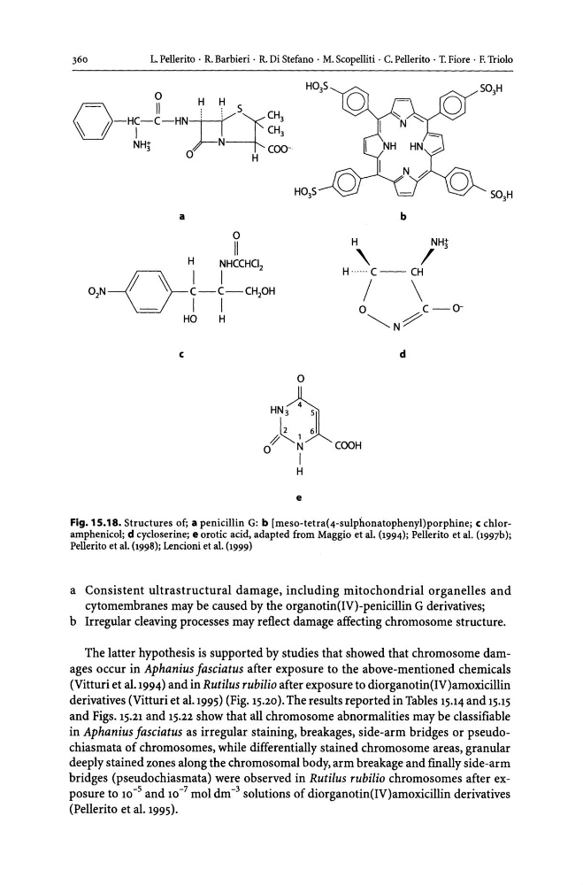
Fig. 15.18. Structures of; a penicillin G: b [meso-tetra(4-sulphonatophenyl)porphine; c chloramphenicol; d cycloserine; e orotic acid, adapted from Maggio et aI. (1994); Pellerito et aI. (1997b);
Pellerito et aI. (1998); Lencioni et aI. (1999)
a Consistent ultrastructural damage, including mitochondrial organelles and
cytomembranes may be caused by the organotin{IV)-penicillin G derivatives;
b Irregular cleaving processes may reflect damage affecting chromosome structure.
The latter hypothesis is supported by studies that showed that chromosome damages occur in Aphanius fasciatus after exposure to the above-mentioned chemicals
(Vitturi et al.1994) and in Rutilus rubilio after exposure to diorganotin(IV)amoxicillin
derivatives (Vitturi et al.1995) (Fig. 15.20). The results reported in Tables 15.14 and 15.15
and Figs. 15.21 and 15.22 show that all chromosome abnormalities may be classifiable
in Aphanius fasciatus as irregular staining, breakages, side-arm bridges or pseudochiasmata of chromosomes, while differentially stained chromosome areas, granular
deeply stained zones along the chromosomal body, arm breakage and finally side-arm
bridges {pseudo chiasmata) were observed in Rutilus rubilio chromosomes after exposure to 10- 5 and 10- 7 mol dm -3 solutions of diorganotin{IV)amoxicillin derivatives
(Pellerito et al. 1995).
L. Pellerito . R. Barbieri· R. Di Stefano . M. Scopelliti . C. Pellerito . T. Fiore . F. Triolo
o
H H
-
II
: ! S
CH 3
O-"y-:C-HNJf~~
NH3
0
H
H0 3 S
S03H
a
b
o
H
I I
02N-o-~ I tCCHCl2
C-C
_
I ICHPH
HO
H
H
NW
' \
/
3
H .... ··C-CH
/ \
o
C-O~N~
c
d
o
tl
HN3
SI
,f'~ 1 6 COOH
o
N
I
H
e
Fig. 15.18. Structures of; a penicillin G: b [meso-tetra(4-sulphonatophenyl)porphine; c chloramphenicol; d cycloserine; e orotic acid, adapted from Maggio et aI. (1994); Pellerito et aI. (1997b);
Pellerito et aI. (1998); Lencioni et aI. (1999)
a Consistent ultrastructural damage, including mitochondrial organelles and
cytomembranes may be caused by the organotin{IV)-penicillin G derivatives;
b Irregular cleaving processes may reflect damage affecting chromosome structure.
The latter hypothesis is supported by studies that showed that chromosome damages occur in Aphanius fasciatus after exposure to the above-mentioned chemicals
(Vitturi et al.1994) and in Rutilus rubilio after exposure to diorganotin(IV)amoxicillin
derivatives (Vitturi et al.1995) (Fig. 15.20). The results reported in Tables 15.14 and 15.15
and Figs. 15.21 and 15.22 show that all chromosome abnormalities may be classifiable
in Aphanius fasciatus as irregular staining, breakages, side-arm bridges or pseudochiasmata of chromosomes, while differentially stained chromosome areas, granular
deeply stained zones along the chromosomal body, arm breakage and finally side-arm
bridges {pseudo chiasmata) were observed in Rutilus rubilio chromosomes after exposure to 10- 5 and 10- 7 mol dm -3 solutions of diorganotin{IV)amoxicillin derivatives
(Pellerito et al. 1995).