

CHAPTER 15 . Toxic Effects of Organometallic Compounds towards Marine Biota
359
In vivo observations showed that cleavage of fertilized eggs was inhibited and embryo blastomeres gave rise to cellular masses not delimited by plasma membranes.
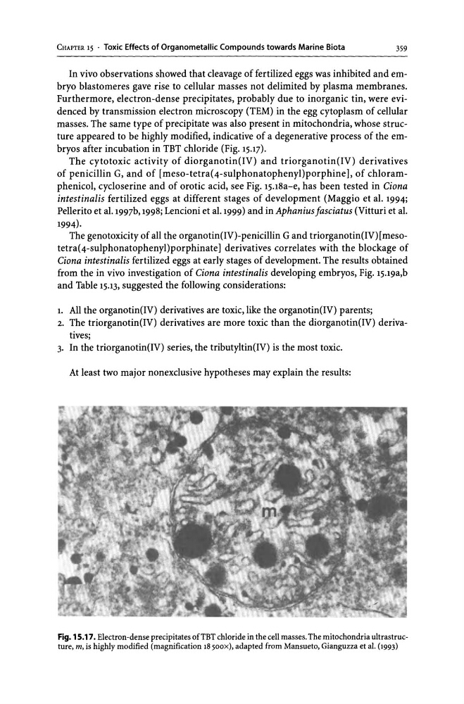
Furthermore, electron-dense precipitates, probably due to inorganic tin, were evidenced by transmission electron microscopy (TEM) in the egg cytoplasm of cellular
masses. The same type of precipitate was also present in mitochondria, whose structure appeared to be highly modified, indicative of a degenerative process of the embryos after incubation in TBT chloride (Fig. 15.17).
The cytotoxic activity of diorganotin(IV) and triorganotin(IV) derivatives
of penicillin G, and of [meso-tetra(4-sulphonatophenyl)porphine], of chloramphenicol, cycloserine and of orotic acid, see Fig. 15.18a-e, has been tested in Ciona
intestinalis fertilized eggs at different stages of development (Maggio et al. 1994;
Pellerito et al.1997b, 1998; Lencioni et al.1999) and in Aphanius Jasciatus (Vitturi et al.
1994).
The genotoxicity of all the organotin(IV)-penicillin G and triorganotin(IV)[mesotetra(4-sulphonatophenyl)porphinate] derivatives correlates with the blockage of
Ciona intestinalis fertilized eggs at early stages of development. The results obtained
from the in vivo investigation of Ciona intestinalis developing embryos, Fig. 15.19a,b
and Table 15.13, suggested the following considerations:
1. All the organotin(IV) derivatives are toxic, like the organotin(IV) parents;
2. The triorganotin(IV) derivatives are more toxic than the diorganotin(IV) derivatives;
3. In the triorganotin(IV) series, the tributyltin(IV) is the most toxic.
At least two major nonexclusive hypotheses may explain the results:
Fig. 15.17. Electron-dense precipitates ofTBT chloride in the cell masses. The mitochondria ultrastructure, m, is highly modified (magnification 18 500X), adapted from Mansueto, Gianguzza et aI. (1993)
359
In vivo observations showed that cleavage of fertilized eggs was inhibited and embryo blastomeres gave rise to cellular masses not delimited by plasma membranes.
Furthermore, electron-dense precipitates, probably due to inorganic tin, were evidenced by transmission electron microscopy (TEM) in the egg cytoplasm of cellular
masses. The same type of precipitate was also present in mitochondria, whose structure appeared to be highly modified, indicative of a degenerative process of the embryos after incubation in TBT chloride (Fig. 15.17).
The cytotoxic activity of diorganotin(IV) and triorganotin(IV) derivatives
of penicillin G, and of [meso-tetra(4-sulphonatophenyl)porphine], of chloramphenicol, cycloserine and of orotic acid, see Fig. 15.18a-e, has been tested in Ciona
intestinalis fertilized eggs at different stages of development (Maggio et al. 1994;
Pellerito et al.1997b, 1998; Lencioni et al.1999) and in Aphanius Jasciatus (Vitturi et al.
1994).
The genotoxicity of all the organotin(IV)-penicillin G and triorganotin(IV)[mesotetra(4-sulphonatophenyl)porphinate] derivatives correlates with the blockage of
Ciona intestinalis fertilized eggs at early stages of development. The results obtained
from the in vivo investigation of Ciona intestinalis developing embryos, Fig. 15.19a,b
and Table 15.13, suggested the following considerations:
1. All the organotin(IV) derivatives are toxic, like the organotin(IV) parents;
2. The triorganotin(IV) derivatives are more toxic than the diorganotin(IV) derivatives;
3. In the triorganotin(IV) series, the tributyltin(IV) is the most toxic.
At least two major nonexclusive hypotheses may explain the results:
Fig. 15.17. Electron-dense precipitates ofTBT chloride in the cell masses. The mitochondria ultrastructure, m, is highly modified (magnification 18 500X), adapted from Mansueto, Gianguzza et aI. (1993)