

35
Chapter two: Oxygen stores and diving
of diving behavior and foraging ecology. This widespread application of the ADL concept
has been based on two observations from Kooyman’s original papers. First, those authors
noted that about 95% of dives were less than the blood lactate-determined ADL of the
Weddell seal. Consequently, a behavioral ADL (ADL b ) has often been determined on the
basis of dive duration distribution with a 90%–95% cutoff threshold for the ADL b .
Kooyman and co-workers also noted that the blood lactate-determined ADL could be
estimated by dividing O 2 stores by a diving metabolic rate (a calculated aerobic dive limit,
ADL c ). The diving metabolic rate was the average metabolic rate measured from O 2 consumption measurements made when the seal surfaced and breathed under a metabolic
dome (Castellini et al. 1992; Ponganis et al. 1993). That metabolic rate was calculated as
the total O 2 consumed during the surface interval divided by the sum of the dive duration and the surface interval. Thus, the metabolic rate in the calculation was not the actual
O 2 consumption rate during a dive, but rather the O 2 consumption rate of the entire dive
event (dive + post-dive surface interval). As emphasized in many reviews (Kooyman and
Ponganis 1998; Ponganis et al. 2011; Ponganis 2015), this calculation does not reflect the
actual status of the body O 2 store at the ADL. It is a simple prediction of the dive duration
associated with the onset of post-dive blood lactate accumulation. The body O 2 store is not
completely depleted at the ADL. The ADL is considered primarily secondary to muscle O 2
depletion in the primary locomotory muscles, with subsequent glycolysis and muscle lactate accumulation during the dive, and lactate washout into blood after the dive. This has
been most completely demonstrated in an avian diver, the emperor penguin (Aptenodytes
forsteri) (Ponganis et al. 1997b; Meir and Ponganis 2009; Williams et al. 2011).
Heart rate (beats per min)
Depth (m)
160
140
120
100
80
60
40
20
0
160
140
120
100
80
60
40
20
0
1
3
5
7
9
1
3
5
Time (min)
7
9
500
400
300
200
100
0
500
400
300
200
100
0
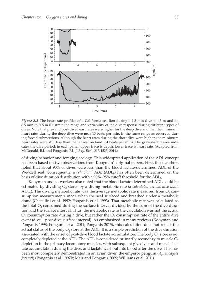
Figure 2.2 The heart rate profiles of a California sea lion during a 1.3 min dive to 45 m and an
8.5 min to 305 m illustrate the range and variability of the dive response during different types of
dives. Note that pre- and post-dive heart rates were higher for the deep dive and that the minimum
heart rates during the deep dive were near 10 beats per min, in the same range as observed during forced submersions. Although the heart rates during the short dive were higher, the minimum
heart rates were still less than that at rest on land (54 beats per min). The gray-shaded area indicates the dive period; in each panel, upper trace is depth, lower trace is heart rate. (Adapted from
McDonald, B.I. and Ponganis, P.J., J. Exp. Biol., 217, 1525, 2014.)
Chapter two: Oxygen stores and diving
of diving behavior and foraging ecology. This widespread application of the ADL concept
has been based on two observations from Kooyman’s original papers. First, those authors
noted that about 95% of dives were less than the blood lactate-determined ADL of the
Weddell seal. Consequently, a behavioral ADL (ADL b ) has often been determined on the
basis of dive duration distribution with a 90%–95% cutoff threshold for the ADL b .
Kooyman and co-workers also noted that the blood lactate-determined ADL could be
estimated by dividing O 2 stores by a diving metabolic rate (a calculated aerobic dive limit,
ADL c ). The diving metabolic rate was the average metabolic rate measured from O 2 consumption measurements made when the seal surfaced and breathed under a metabolic
dome (Castellini et al. 1992; Ponganis et al. 1993). That metabolic rate was calculated as
the total O 2 consumed during the surface interval divided by the sum of the dive duration and the surface interval. Thus, the metabolic rate in the calculation was not the actual
O 2 consumption rate during a dive, but rather the O 2 consumption rate of the entire dive
event (dive + post-dive surface interval). As emphasized in many reviews (Kooyman and
Ponganis 1998; Ponganis et al. 2011; Ponganis 2015), this calculation does not reflect the
actual status of the body O 2 store at the ADL. It is a simple prediction of the dive duration
associated with the onset of post-dive blood lactate accumulation. The body O 2 store is not
completely depleted at the ADL. The ADL is considered primarily secondary to muscle O 2
depletion in the primary locomotory muscles, with subsequent glycolysis and muscle lactate accumulation during the dive, and lactate washout into blood after the dive. This has
been most completely demonstrated in an avian diver, the emperor penguin (Aptenodytes
forsteri) (Ponganis et al. 1997b; Meir and Ponganis 2009; Williams et al. 2011).
Heart rate (beats per min)
Depth (m)
160
140
120
100
80
60
40
20
0
160
140
120
100
80
60
40
20
0
1
3
5
7
9
1
3
5
Time (min)
7
9
500
400
300
200
100
0
500
400
300
200
100
0
Figure 2.2 The heart rate profiles of a California sea lion during a 1.3 min dive to 45 m and an
8.5 min to 305 m illustrate the range and variability of the dive response during different types of
dives. Note that pre- and post-dive heart rates were higher for the deep dive and that the minimum
heart rates during the deep dive were near 10 beats per min, in the same range as observed during forced submersions. Although the heart rates during the short dive were higher, the minimum
heart rates were still less than that at rest on land (54 beats per min). The gray-shaded area indicates the dive period; in each panel, upper trace is depth, lower trace is heart rate. (Adapted from
McDonald, B.I. and Ponganis, P.J., J. Exp. Biol., 217, 1525, 2014.)