

(a)
(b)
Figure 6.2 (a) Pygmy blue whale, feeding. Showing baleen, Portland, Victoria, Australia. (Photo by
Paul Enser, Australian Antarctic Division, Kingston, South Australia, Australia.) (b) Crabeater seal
head and shoulders illustrating the heavily cusped post-canine teeth used by this species to sieve
Antarctic krill. (Photo by Mark A. Hindell.)
Figure 6.3 Direct observation of sub-surface feeding events is rare. Sub-surface feeding on bottomdwelling or mid-water prey, such as squid as shown here, are greatly underestimated in this method
(Chiroteuthis sp.). (Photo by Glenn Jacobson, Australian Antarctic Division © Commonwealth of
Australia.)
Urine
1. Glomerulus
2. Proximal convoluted tubule
3. Proximal straight tubule
4. Descending thin limb of loop of Henle
5. Ascending thin limb of loop of Henle
6. Thick ascending limb of loop of Henle
7. Distal convoluted tubule
8. Connecting tubule
9. Collecting duct
Na
+ , K
+
Water
urea
Na + , K +
Ca 2+
Na + , Ca 2+
Water
Higher urea
concentration
in the interstitium
Inner medulla Outer medulla Cortex
Water
Inner
stripe
Outer
stripe
Blood flow
Filtered
plasma
5
6
2
1
9
8
7
4
3
A histological section of the renicule
of a bottlenose dolphin
Cortex
Renicule
(a)
(b)
Kidney
Renal
papilla
Medulla
Glucose
urea
water
Na + , Cl –
K + , Ca 2+
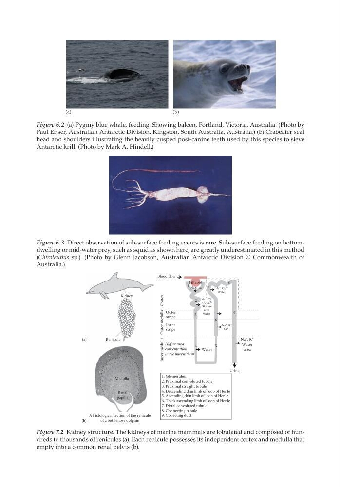
Figure 7.2 Kidney structure. The kidneys of marine mammals are lobulated and composed of hundreds to thousands of renicules (a). Each renicule possesses its independent cortex and medulla that
empty into a common renal pelvis (b).
(b)
Figure 6.2 (a) Pygmy blue whale, feeding. Showing baleen, Portland, Victoria, Australia. (Photo by
Paul Enser, Australian Antarctic Division, Kingston, South Australia, Australia.) (b) Crabeater seal
head and shoulders illustrating the heavily cusped post-canine teeth used by this species to sieve
Antarctic krill. (Photo by Mark A. Hindell.)
Figure 6.3 Direct observation of sub-surface feeding events is rare. Sub-surface feeding on bottomdwelling or mid-water prey, such as squid as shown here, are greatly underestimated in this method
(Chiroteuthis sp.). (Photo by Glenn Jacobson, Australian Antarctic Division © Commonwealth of
Australia.)
Urine
1. Glomerulus
2. Proximal convoluted tubule
3. Proximal straight tubule
4. Descending thin limb of loop of Henle
5. Ascending thin limb of loop of Henle
6. Thick ascending limb of loop of Henle
7. Distal convoluted tubule
8. Connecting tubule
9. Collecting duct
Na
+ , K
+
Water
urea
Na + , K +
Ca 2+
Na + , Ca 2+
Water
Higher urea
concentration
in the interstitium
Inner medulla Outer medulla Cortex
Water
Inner
stripe
Outer
stripe
Blood flow
Filtered
plasma
5
6
2
1
9
8
7
4
3
A histological section of the renicule
of a bottlenose dolphin
Cortex
Renicule
(a)
(b)
Kidney
Renal
papilla
Medulla
Glucose
urea
water
Na + , Cl –
K + , Ca 2+
Figure 7.2 Kidney structure. The kidneys of marine mammals are lobulated and composed of hundreds to thousands of renicules (a). Each renicule possesses its independent cortex and medulla that
empty into a common renal pelvis (b).