

Ontogeny of Marine Turtle Gonads
215
7.6.2 TESTES
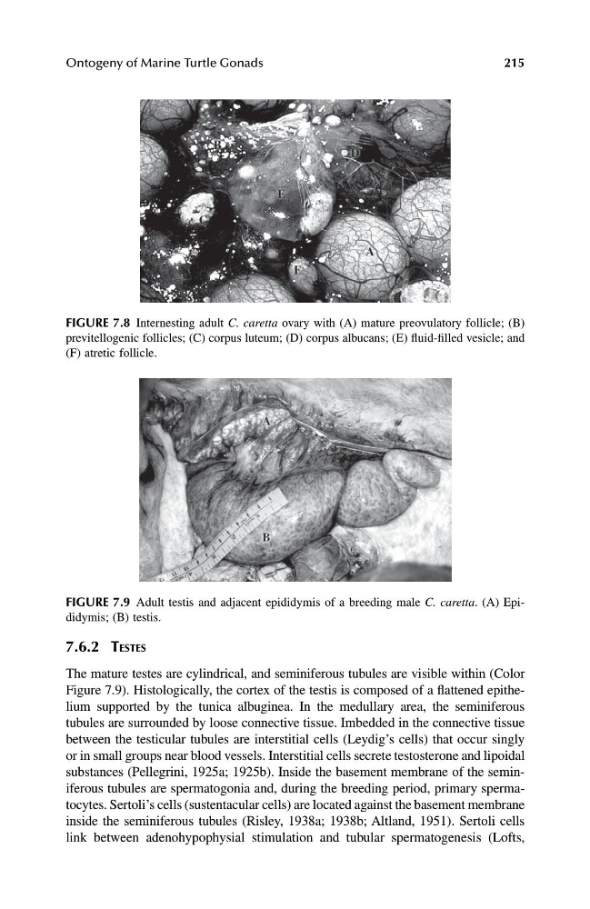
The mature testes are cylindrical, and seminiferous tubules are visible within (Color
Figure 7.9). Histologically, the cortex of the testis is composed of a flattened epithelium supported by the tunica albuginea. In the medullary area, the seminiferous
tubules are surrounded by loose connective tissue. Imbedded in the connective tissue
between the testicular tubules are interstitial cells (Leydig’s cells) that occur singly
or in small groups near blood vessels. Interstitial cells secrete testosterone and lipoidal
substances (Pellegrini, 1925a; 1925b). Inside the basement membrane of the seminiferous tubules are spermatogonia and, during the breeding period, primary spermatocytes. Sertoli’s cells (sustentacular cells) are located against the basement membrane
inside the seminiferous tubules (Risley, 1938a; 1938b; Altland, 1951). Sertoli cells
link between adenohypophysial stimulation and tubular spermatogenesis (Lofts,
FIGURE 7.8 Internesting adult C. caretta ovary with (A) mature preovulatory follicle; (B)
previtellogenic follicles; (C) corpus luteum; (D) corpus albucans; (E) fluid-filled vesicle; and
(F) atretic follicle.
FIGURE 7.9 Adult testis and adjacent epididymis of a breeding male C. caretta. (A) Epididymis; (B) testis.
1123 book.book Page 215 Monday, November 11, 2002 11:11 AM
215
7.6.2 TESTES
The mature testes are cylindrical, and seminiferous tubules are visible within (Color
Figure 7.9). Histologically, the cortex of the testis is composed of a flattened epithelium supported by the tunica albuginea. In the medullary area, the seminiferous
tubules are surrounded by loose connective tissue. Imbedded in the connective tissue
between the testicular tubules are interstitial cells (Leydig’s cells) that occur singly
or in small groups near blood vessels. Interstitial cells secrete testosterone and lipoidal
substances (Pellegrini, 1925a; 1925b). Inside the basement membrane of the seminiferous tubules are spermatogonia and, during the breeding period, primary spermatocytes. Sertoli’s cells (sustentacular cells) are located against the basement membrane
inside the seminiferous tubules (Risley, 1938a; 1938b; Altland, 1951). Sertoli cells
link between adenohypophysial stimulation and tubular spermatogenesis (Lofts,
FIGURE 7.8 Internesting adult C. caretta ovary with (A) mature preovulatory follicle; (B)
previtellogenic follicles; (C) corpus luteum; (D) corpus albucans; (E) fluid-filled vesicle; and
(F) atretic follicle.
FIGURE 7.9 Adult testis and adjacent epididymis of a breeding male C. caretta. (A) Epididymis; (B) testis.
1123 book.book Page 215 Monday, November 11, 2002 11:11 AM