

The External, Musculoskeletal and Neuro-Anatomy of Sea Turtles
71
2.5.2 CENTRAL NERVOUS SYSTEM
Traditionally, the brain (Figure 2.26) is described by three regions that arise as
distinct vesicles during development: the forebrain, midbrain, and hindbrain. The
following combinations of external and internal landmarks roughly identify these
divisions. The forebrain extends from the nose to the posterior cerebrum. The
midbrain extends from the eye to the posterior aspect of the optic lobes. The
hindbrain extends from the ear to the posterior cerebellum (Romer and Parsons,
1986). These regions, in turn, are subdivided topographically and/or histochemically into principal divisions: the telencephalon and diencephalon of the forebrain,
the mesencephalon of the midbrain, and the metencephalon and myelencephalon
of the hindbrain (Hyman and Wake, 1979).
The divisions of the brain (Figures 2.26 and 2.27) and their major components
are as follows:
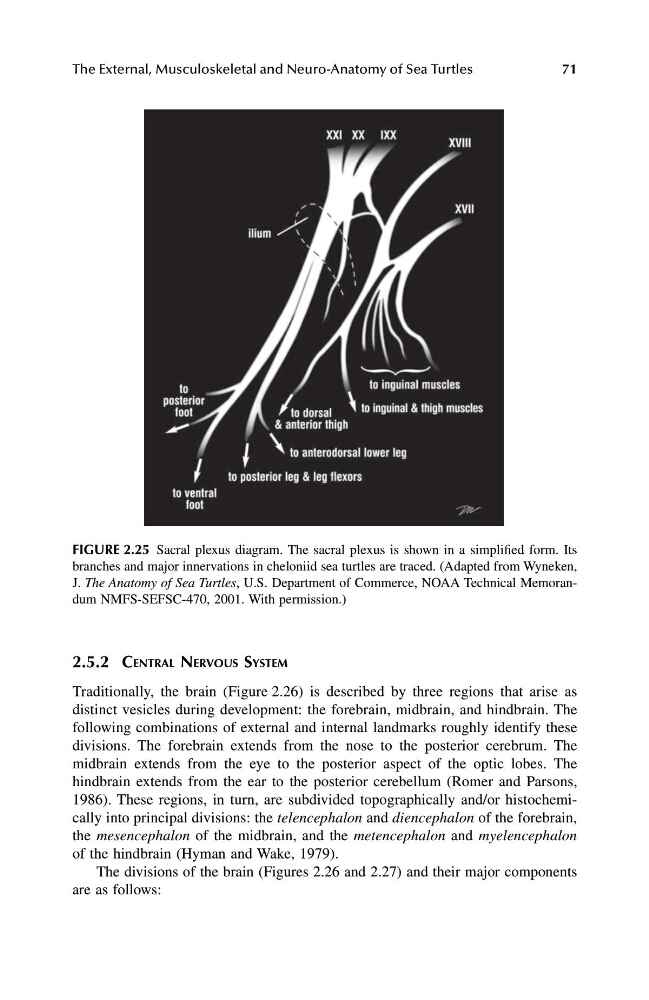
FIGURE 2.25 Sacral plexus diagram. The sacral plexus is shown in a simplified form. Its
branches and major innervations in cheloniid sea turtles are traced. (Adapted from Wyneken,
J. The Anatomy of Sea Turtles, U.S. Department of Commerce, NOAA Technical Memorandum NMFS-SEFSC-470, 2001. With permission.)
1123_C02.fm Page 71 Friday, November 15, 2002 11:16 AM
71
2.5.2 CENTRAL NERVOUS SYSTEM
Traditionally, the brain (Figure 2.26) is described by three regions that arise as
distinct vesicles during development: the forebrain, midbrain, and hindbrain. The
following combinations of external and internal landmarks roughly identify these
divisions. The forebrain extends from the nose to the posterior cerebrum. The
midbrain extends from the eye to the posterior aspect of the optic lobes. The
hindbrain extends from the ear to the posterior cerebellum (Romer and Parsons,
1986). These regions, in turn, are subdivided topographically and/or histochemically into principal divisions: the telencephalon and diencephalon of the forebrain,
the mesencephalon of the midbrain, and the metencephalon and myelencephalon
of the hindbrain (Hyman and Wake, 1979).
The divisions of the brain (Figures 2.26 and 2.27) and their major components
are as follows:
FIGURE 2.25 Sacral plexus diagram. The sacral plexus is shown in a simplified form. Its
branches and major innervations in cheloniid sea turtles are traced. (Adapted from Wyneken,
J. The Anatomy of Sea Turtles, U.S. Department of Commerce, NOAA Technical Memorandum NMFS-SEFSC-470, 2001. With permission.)
1123_C02.fm Page 71 Friday, November 15, 2002 11:16 AM