

70
Biology of Sea Turtles, Vol. II
The sacral plexus (Figure 2.25) arises as four (sometimes five to six) branches
from spinal nerves XVII–XXI associated with the last trunk vertebra and the sacral
vertebrae. The sacral plexus is poorly studied in turtles and described only grossly
in sea turtles (Walker, 1973; Wyneken, 2001). These nerves interconnect and
subdivide several times to send nerves to the inguinal, pelvic, and hind leg muscles
(Table 2.2). The more posterior nerve roots give rise to the obturator nerve, which
goes to the ventral pelvic muscles, and the ichiadicus nerve, which runs medial
to the ilium and then divides to form the peroneal and sciatic nerves (Figure 2.25).
The anterior two nerves interconnect to provide major innervations (via crural,
femoral, and tibial nerves) to the inguinal muscles, thigh adductors, and leg
extensors (Figure 2.25).
Like those nerves forming the cervical plexus, these sacral nerves arise in dorsal
and ventral pairs, with the dorsal nerves generally innervating dorsal hind limb
muscles and ventral nerves generally innervating ventral hind limb muscles. However, many pelvic and hind limb muscles receive multiple innervations. Hence, it is
common for muscles to have innervations from multiple spinal nerves and have
branches from both dorsal and ventral nerve tracks going to the same muscle.
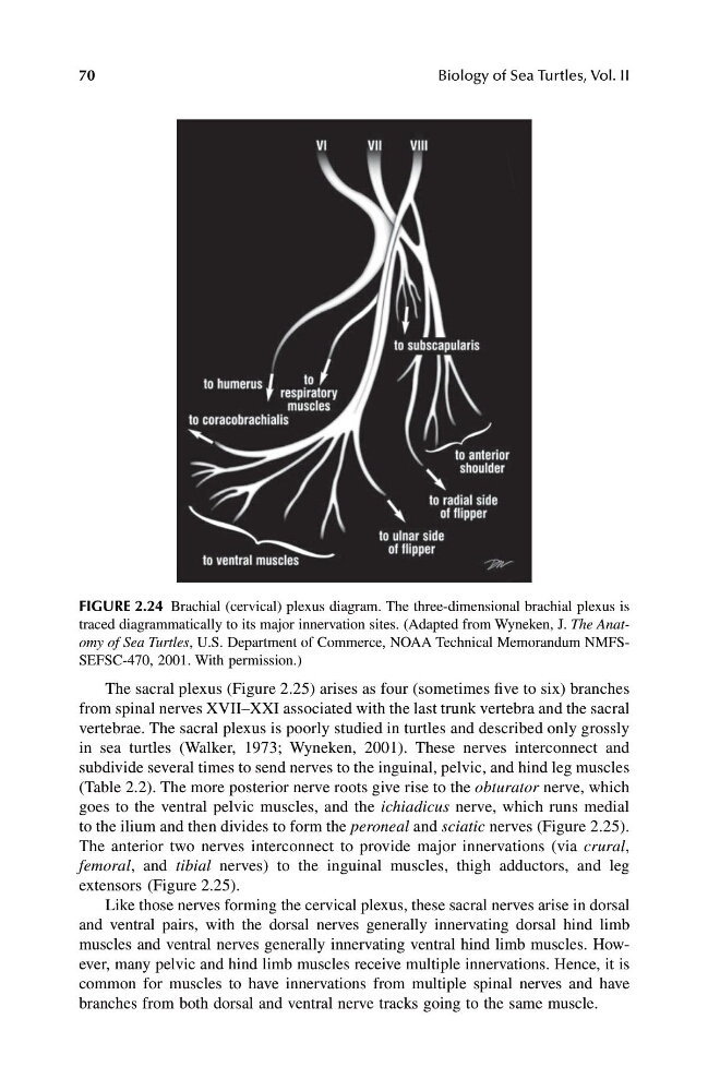
FIGURE 2.24 Brachial (cervical) plexus diagram. The three-dimensional brachial plexus is
traced diagrammatically to its major innervation sites. (Adapted from Wyneken, J. The Anatomy of Sea Turtles, U.S. Department of Commerce, NOAA Technical Memorandum NMFSSEFSC-470, 2001. With permission.)
1123_C02.fm Page 70 Friday, November 15, 2002 11:16 AM
Biology of Sea Turtles, Vol. II
The sacral plexus (Figure 2.25) arises as four (sometimes five to six) branches
from spinal nerves XVII–XXI associated with the last trunk vertebra and the sacral
vertebrae. The sacral plexus is poorly studied in turtles and described only grossly
in sea turtles (Walker, 1973; Wyneken, 2001). These nerves interconnect and
subdivide several times to send nerves to the inguinal, pelvic, and hind leg muscles
(Table 2.2). The more posterior nerve roots give rise to the obturator nerve, which
goes to the ventral pelvic muscles, and the ichiadicus nerve, which runs medial
to the ilium and then divides to form the peroneal and sciatic nerves (Figure 2.25).
The anterior two nerves interconnect to provide major innervations (via crural,
femoral, and tibial nerves) to the inguinal muscles, thigh adductors, and leg
extensors (Figure 2.25).
Like those nerves forming the cervical plexus, these sacral nerves arise in dorsal
and ventral pairs, with the dorsal nerves generally innervating dorsal hind limb
muscles and ventral nerves generally innervating ventral hind limb muscles. However, many pelvic and hind limb muscles receive multiple innervations. Hence, it is
common for muscles to have innervations from multiple spinal nerves and have
branches from both dorsal and ventral nerve tracks going to the same muscle.
FIGURE 2.24 Brachial (cervical) plexus diagram. The three-dimensional brachial plexus is
traced diagrammatically to its major innervation sites. (Adapted from Wyneken, J. The Anatomy of Sea Turtles, U.S. Department of Commerce, NOAA Technical Memorandum NMFSSEFSC-470, 2001. With permission.)
1123_C02.fm Page 70 Friday, November 15, 2002 11:16 AM