

(4) Restricted fragment length polymorphisms (RFLP)
Mitochondrial DNA D-loop region polymorph (Fig. 5.4): All DNA, including
mitochrondrial DNA, is extracted and the target region is amplified through PCR.
After restriction enzyme truncation, DNA fragments are separated by electrophoresis, dyed with ethidium bromide, and exposed to UV rays (with a transilluminator) to be recorded by Polaroid camera or other means. Because this
approach uses PCR, no radioactive isotope is needed, and polymorph detection can
be carried out simply in an ordinary biological laboratory without special facilities
or equipment (Fig. 5.4; Ishida et al. 1994).
For a PCR primer, a base sequence is designed so that the D-loop region is
effectively embedded in the center.
Amplified fragment length polymorphisms (AFLP): A small quantity of nuclear
DNA is truncated with restriction enzymes and an adapter sequence is attached to
the fragment end to serve as a primer. The primer, which includes the restriction
enzyme recognition sequence, is used for PCR processing. Because so many
amplified fragments are present, PCR is conducted twice using a primer including a
random (facultative) base. Placement of DNA size markers at either end during
electrophoresis allows for easy identification of bands and comparison between
different films. This is an excellent technology that combines the reproducibility of
RFLP with the simplicity of PCR, allowing for processing of multiple samples.
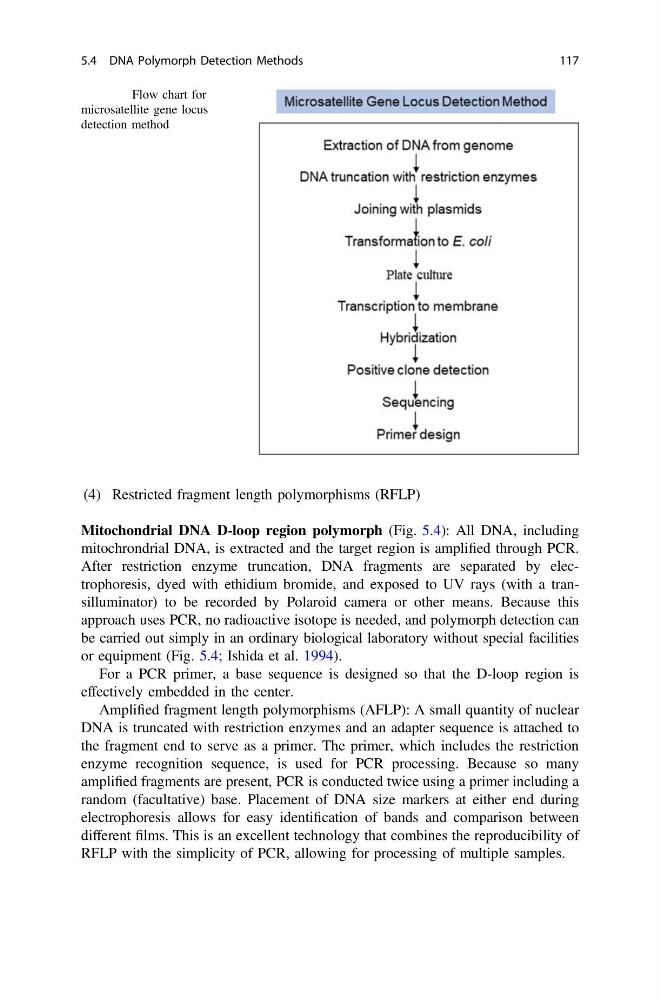
Fig. 5.3 Flow chart for
microsatellite gene locus
detection method
5.4 DNA Polymorph Detection Methods
117
Mitochondrial DNA D-loop region polymorph (Fig. 5.4): All DNA, including
mitochrondrial DNA, is extracted and the target region is amplified through PCR.
After restriction enzyme truncation, DNA fragments are separated by electrophoresis, dyed with ethidium bromide, and exposed to UV rays (with a transilluminator) to be recorded by Polaroid camera or other means. Because this
approach uses PCR, no radioactive isotope is needed, and polymorph detection can
be carried out simply in an ordinary biological laboratory without special facilities
or equipment (Fig. 5.4; Ishida et al. 1994).
For a PCR primer, a base sequence is designed so that the D-loop region is
effectively embedded in the center.
Amplified fragment length polymorphisms (AFLP): A small quantity of nuclear
DNA is truncated with restriction enzymes and an adapter sequence is attached to
the fragment end to serve as a primer. The primer, which includes the restriction
enzyme recognition sequence, is used for PCR processing. Because so many
amplified fragments are present, PCR is conducted twice using a primer including a
random (facultative) base. Placement of DNA size markers at either end during
electrophoresis allows for easy identification of bands and comparison between
different films. This is an excellent technology that combines the reproducibility of
RFLP with the simplicity of PCR, allowing for processing of multiple samples.
Fig. 5.3 Flow chart for
microsatellite gene locus
detection method
5.4 DNA Polymorph Detection Methods
117