

The Great Barrier Reef
134
producers, grazers, nitrogen fixers and predators)
(Box 14.2); macroplankton (20–200 mm) (larvaceans, larval fishes and other zooplankton that are grazers and
predators) and megaplankton (over 200 mm) (jellyfish,
salps and other zooplankton that are mainly grazer and
predators).
Water bottle samples (organisms <50 microns)
Our primary focus here is organisms between 2 μm and
20 μm in size: the nanoplankton. Members of the nanoplankton are very small and characterised by a high surface area to volume ratio. The surface area to volume
ratio influences the amounts of materials exchanged
with the environment. In general, an inverse relationship between size and reproductive capacity is observed
(i.e. small cells have a much higher reproductive rate
than large cells), although this relationship is not easily
applied to flagellated forms of the different size classes
and some diatoms can outgrow picoplankton (e.g.
Prochlorococcus). The high reproductive capacity is linked
to the ability to immediately respond to short nutrient
pulses. Therefore, nanoplankton often occurs at high
biomass per unit volume. For example, high biomass
patches can stretch over kilometres and the resultant
chlorophyll fluorescence is readily visible from space via
satellites. These population/community maxima may,
however, be short-lived and can change dramatically
within the space of a day or two. Grazing is another
factor that controls the biomass of these very small
organisms, as they are readily consumed by grazers such
as protozoa, pelagic tunicates, rotifers and crustaceans.
Phytoplankton of this size range may occur in colonies and as flagellated and non-flagellated single cells,
which are often very delicate and generally do not preserve well. In particular, flagellated forms tend to loose
their flagella and shape upon fixation with either
Lugol’s solution or formaldehyde. In most cases, the
presence/absence and number of flagellae, as well as
swimming behaviour are essential criteria for the
identification to genus and species. Due to the miniscule size of these organisms, swimming behaviour and
patterns of flagellation of live material must be
observed at high magnifications (400 and more)
requiring special contrasting objectives to visualise
these structures (i.e. differential interference contrast
or phase contrast). Even if swimming behaviour can
be observed in live samples, the presence and number
of flagellae typically remains a mystery as these
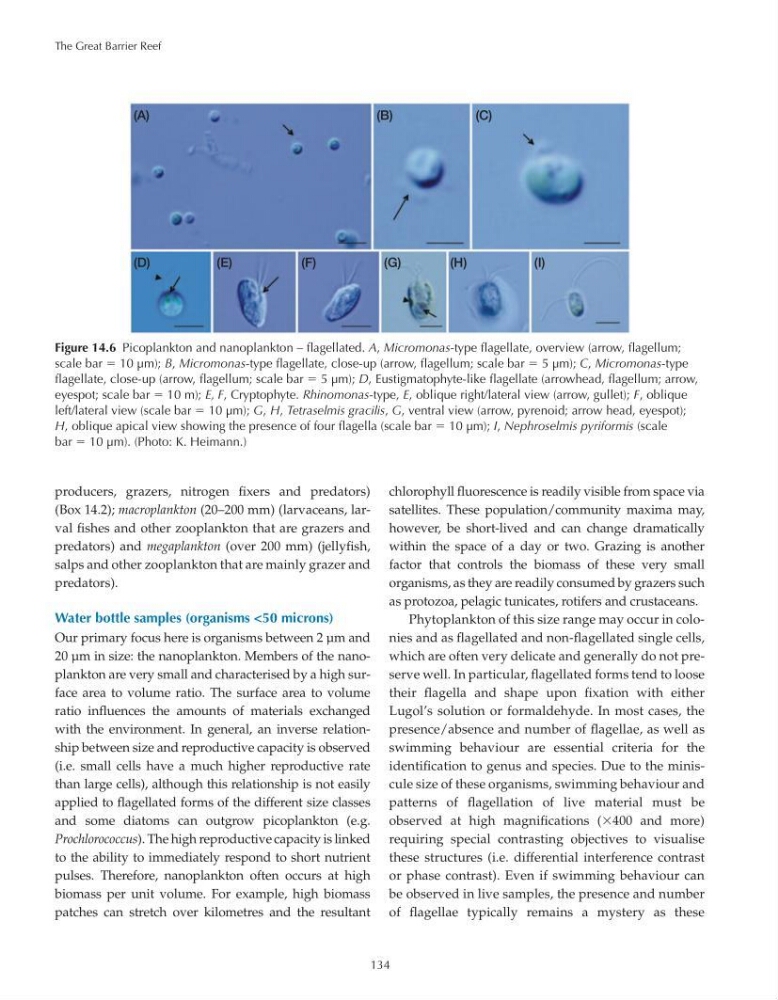
Figure 14.6 Picoplankton and nanoplankton – flagellated. A, Micromonas-type flagellate, overview (arrow, flagellum;
scale bar 10 μm); B, Micromonas-type flagellate, close-up (arrow, flagellum; scale bar 5 μm); C, Micromonas-type
flagellate, close-up (arrow, flagellum; scale bar 5 μm); D, Eustigmatophyte-like flagellate (arrowhead, flagellum; arrow,
eyespot; scale bar 10 m); E, F, Cryptophyte. Rhinomonas-type, E, oblique right/lateral view (arrow, gullet); F, oblique
left/lateral view (scale bar 10 μm); G, H, Tetraselmis gracilis, G, ventral view (arrow, pyrenoid; arrow head, eyespot);
H, oblique apical view showing the presence of four flagella (scale bar 10 μm); I, Nephroselmis pyriformis (scale
bar 10 μm). (Photo: K. Heimann.)
(A)
(D)
(E)
(F)
(G)
(H)
(I)
(B)
(C)
134
producers, grazers, nitrogen fixers and predators)
(Box 14.2); macroplankton (20–200 mm) (larvaceans, larval fishes and other zooplankton that are grazers and
predators) and megaplankton (over 200 mm) (jellyfish,
salps and other zooplankton that are mainly grazer and
predators).
Water bottle samples (organisms <50 microns)
Our primary focus here is organisms between 2 μm and
20 μm in size: the nanoplankton. Members of the nanoplankton are very small and characterised by a high surface area to volume ratio. The surface area to volume
ratio influences the amounts of materials exchanged
with the environment. In general, an inverse relationship between size and reproductive capacity is observed
(i.e. small cells have a much higher reproductive rate
than large cells), although this relationship is not easily
applied to flagellated forms of the different size classes
and some diatoms can outgrow picoplankton (e.g.
Prochlorococcus). The high reproductive capacity is linked
to the ability to immediately respond to short nutrient
pulses. Therefore, nanoplankton often occurs at high
biomass per unit volume. For example, high biomass
patches can stretch over kilometres and the resultant
chlorophyll fluorescence is readily visible from space via
satellites. These population/community maxima may,
however, be short-lived and can change dramatically
within the space of a day or two. Grazing is another
factor that controls the biomass of these very small
organisms, as they are readily consumed by grazers such
as protozoa, pelagic tunicates, rotifers and crustaceans.
Phytoplankton of this size range may occur in colonies and as flagellated and non-flagellated single cells,
which are often very delicate and generally do not preserve well. In particular, flagellated forms tend to loose
their flagella and shape upon fixation with either
Lugol’s solution or formaldehyde. In most cases, the
presence/absence and number of flagellae, as well as
swimming behaviour are essential criteria for the
identification to genus and species. Due to the miniscule size of these organisms, swimming behaviour and
patterns of flagellation of live material must be
observed at high magnifications (400 and more)
requiring special contrasting objectives to visualise
these structures (i.e. differential interference contrast
or phase contrast). Even if swimming behaviour can
be observed in live samples, the presence and number
of flagellae typically remains a mystery as these
Figure 14.6 Picoplankton and nanoplankton – flagellated. A, Micromonas-type flagellate, overview (arrow, flagellum;
scale bar 10 μm); B, Micromonas-type flagellate, close-up (arrow, flagellum; scale bar 5 μm); C, Micromonas-type
flagellate, close-up (arrow, flagellum; scale bar 5 μm); D, Eustigmatophyte-like flagellate (arrowhead, flagellum; arrow,
eyespot; scale bar 10 m); E, F, Cryptophyte. Rhinomonas-type, E, oblique right/lateral view (arrow, gullet); F, oblique
left/lateral view (scale bar 10 μm); G, H, Tetraselmis gracilis, G, ventral view (arrow, pyrenoid; arrow head, eyespot);
H, oblique apical view showing the presence of four flagella (scale bar 10 μm); I, Nephroselmis pyriformis (scale
bar 10 μm). (Photo: K. Heimann.)
(A)
(D)
(E)
(F)
(G)
(H)
(I)
(B)
(C)