

72
A. Boudaya et al.
– Signals Annotation
To evaluate each individual’s state of exhaustion, we concentrate on the brain
areas that are responsible for hypo-vigilance detection. In this regard, different brain waves are targeted such as [16]:
• Delta waves refer to consciousness, sleep or deep sleep states. These
waves were found in the temporal and occipital conditions with low frequency (less than 4 Hz) and high amplitude.
• Theta waves design the relaxation and hypnosis states with a range
of frequency between 4 and 8 Hz. Theta waves are extracted from the
temporal zone and are produced during the first phase of slow sleep or in
deep relaxation state.
• Alpha waves refer to waking but relaxed states. These waves are captured in the posterior part, precisely the occipital region, with a frequency
interval between 8 and 12 Hz and a low amplitude interval between 20
and 60 µV.
• Beta waves relate to alertness states. These waves are captured from
the temporal and occipital lobes of the brain. They are characterized by
high frequency interval of 12 to 30 Hz with a low amplitude interval of 10
to 30 µV.
• Gamma waves refer to hypervigilance states with a frequency interval
between 30 to 80 Hz.
In the data annotation step, we only use the O1 and O2 electrodes of occipital
zone which are responsible for drowsiness sensation.
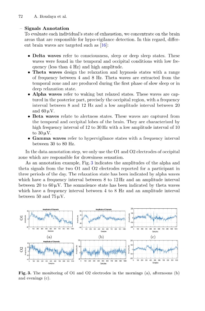
As an annotation example, Fig. 3 indicates the amplitudes of the alpha and
theta signals from the two O1 and O2 electrodes reported for a participant in
three periods of the day. The relaxation state has been indicated by alpha waves
which have a frequency interval between 8 to 12 Hz and an amplitude interval
between 20 to 60 µV. The somnolence state has been indicated by theta waves
which have a frequency interval between 4 to 8 Hz and an amplitude interval
between 50 and 75 µV.
Fig. 3. The monitoring of O1 and O2 electrodes in the mornings (a), afternoons (b)
and evenings (c).
A. Boudaya et al.
– Signals Annotation
To evaluate each individual’s state of exhaustion, we concentrate on the brain
areas that are responsible for hypo-vigilance detection. In this regard, different brain waves are targeted such as [16]:
• Delta waves refer to consciousness, sleep or deep sleep states. These
waves were found in the temporal and occipital conditions with low frequency (less than 4 Hz) and high amplitude.
• Theta waves design the relaxation and hypnosis states with a range
of frequency between 4 and 8 Hz. Theta waves are extracted from the
temporal zone and are produced during the first phase of slow sleep or in
deep relaxation state.
• Alpha waves refer to waking but relaxed states. These waves are captured in the posterior part, precisely the occipital region, with a frequency
interval between 8 and 12 Hz and a low amplitude interval between 20
and 60 µV.
• Beta waves relate to alertness states. These waves are captured from
the temporal and occipital lobes of the brain. They are characterized by
high frequency interval of 12 to 30 Hz with a low amplitude interval of 10
to 30 µV.
• Gamma waves refer to hypervigilance states with a frequency interval
between 30 to 80 Hz.
In the data annotation step, we only use the O1 and O2 electrodes of occipital
zone which are responsible for drowsiness sensation.
As an annotation example, Fig. 3 indicates the amplitudes of the alpha and
theta signals from the two O1 and O2 electrodes reported for a participant in
three periods of the day. The relaxation state has been indicated by alpha waves
which have a frequency interval between 8 to 12 Hz and an amplitude interval
between 20 to 60 µV. The somnolence state has been indicated by theta waves
which have a frequency interval between 4 to 8 Hz and an amplitude interval
between 50 and 75 µV.
Fig. 3. The monitoring of O1 and O2 electrodes in the mornings (a), afternoons (b)
and evenings (c).