

Superpixel Segmentation for CC Parcellation in MRI
121
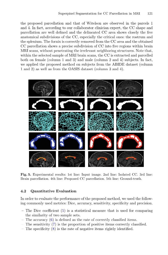
the proposed parcellation and that of Witelson are observed in the parcels 1
and 4. In fact, according to our collaborator clinician expert, the CC shape and
parcellation are well defined and the delineated CC area shows closely the five
anatomical subdivisions of the CC, especially the critical ones: the rostrum and
the splenium. The fornix is correctly removed from the CC area and the obtained
CC parcellation shows a precise subdivision of CC into five regions within brain
MRI scans, without penetrating the irrelevant neighboring structures. Note that,
within the selected sample of MRI brain scans, the CC is extracted and parcelled
both on female (column 1 and 3) and male (column 2 and 4) subjects. In fact,
we applied the proposed method on subjects from the ABIDE dataset (column
1 and 2) as well as from the OASIS dataset (column 3 and 4).
Fig. 5. Experimental results: 1st line: Input image. 2nd line: Isolated CC. 3rd line:
Brain parcellation. 4th line: Proposed CC parcellation. 5th line: Ground-truth.
4.2 Quantitative Evaluation
In order to evaluate the performance of the proposed method, we used the following commonly used metrics: Dice, accuracy, sensitivity, specificity and precision.
– The Dice coefficient (5) is a statistical measure that is used for comparing
the similarity of two sample sets.
– The accuracy (6) is defined as the rate of correctly classified items.
– The sensitivity (7) is the proportion of positive items correctly classified.
– The specificity (8) is the rate of negative items rightly identified.
121
the proposed parcellation and that of Witelson are observed in the parcels 1
and 4. In fact, according to our collaborator clinician expert, the CC shape and
parcellation are well defined and the delineated CC area shows closely the five
anatomical subdivisions of the CC, especially the critical ones: the rostrum and
the splenium. The fornix is correctly removed from the CC area and the obtained
CC parcellation shows a precise subdivision of CC into five regions within brain
MRI scans, without penetrating the irrelevant neighboring structures. Note that,
within the selected sample of MRI brain scans, the CC is extracted and parcelled
both on female (column 1 and 3) and male (column 2 and 4) subjects. In fact,
we applied the proposed method on subjects from the ABIDE dataset (column
1 and 2) as well as from the OASIS dataset (column 3 and 4).
Fig. 5. Experimental results: 1st line: Input image. 2nd line: Isolated CC. 3rd line:
Brain parcellation. 4th line: Proposed CC parcellation. 5th line: Ground-truth.
4.2 Quantitative Evaluation
In order to evaluate the performance of the proposed method, we used the following commonly used metrics: Dice, accuracy, sensitivity, specificity and precision.
– The Dice coefficient (5) is a statistical measure that is used for comparing
the similarity of two sample sets.
– The accuracy (6) is defined as the rate of correctly classified items.
– The sensitivity (7) is the proportion of positive items correctly classified.
– The specificity (8) is the rate of negative items rightly identified.