

120
A. Jlassi et al.
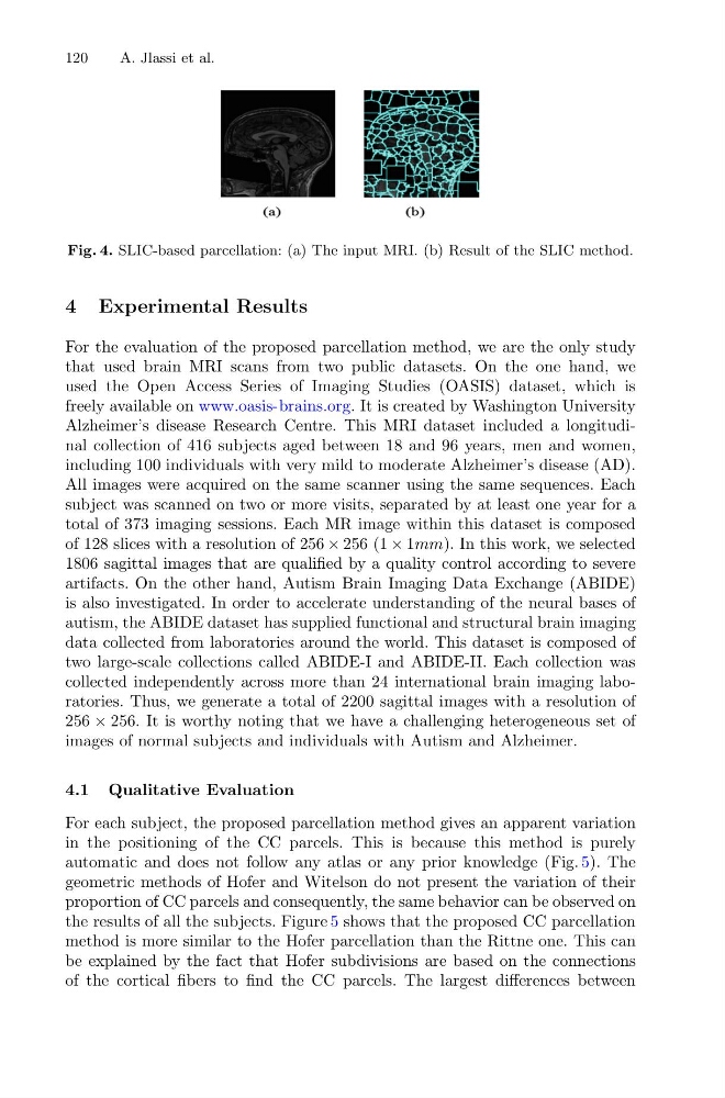
Fig. 4. SLIC-based parcellation: (a) The input MRI. (b) Result of the SLIC method.
4 Experimental Results
For the evaluation of the proposed parcellation method, we are the only study
that used brain MRI scans from two public datasets. On the one hand, we
used the Open Access Series of Imaging Studies (OASIS) dataset, which is
freely available on www.oasis-brains.org. It is created by Washington University
Alzheimer’s disease Research Centre. This MRI dataset included a longitudinal collection of 416 subjects aged between 18 and 96 years, men and women,
including 100 individuals with very mild to moderate Alzheimer’s disease (AD).
All images were acquired on the same scanner using the same sequences. Each
subject was scanned on two or more visits, separated by at least one year for a
total of 373 imaging sessions. Each MR image within this dataset is composed
of 128 slices with a resolution of 256 × 256 (1 × 1mm). In this work, we selected
1806 sagittal images that are qualified by a quality control according to severe
artifacts. On the other hand, Autism Brain Imaging Data Exchange (ABIDE)
is also investigated. In order to accelerate understanding of the neural bases of
autism, the ABIDE dataset has supplied functional and structural brain imaging
data collected from laboratories around the world. This dataset is composed of
two large-scale collections called ABIDE-I and ABIDE-II. Each collection was
collected independently across more than 24 international brain imaging laboratories. Thus, we generate a total of 2200 sagittal images with a resolution of
256 × 256. It is worthy noting that we have a challenging heterogeneous set of
images of normal subjects and individuals with Autism and Alzheimer.
4.1 Qualitative Evaluation
For each subject, the proposed parcellation method gives an apparent variation
in the positioning of the CC parcels. This is because this method is purely
automatic and does not follow any atlas or any prior knowledge (Fig. 5). The
geometric methods of Hofer and Witelson do not present the variation of their
proportion of CC parcels and consequently, the same behavior can be observed on
the results of all the subjects. Figure 5 shows that the proposed CC parcellation
method is more similar to the Hofer parcellation than the Rittne one. This can
be explained by the fact that Hofer subdivisions are based on the connections
of the cortical fibers to find the CC parcels. The largest differences between
A. Jlassi et al.
Fig. 4. SLIC-based parcellation: (a) The input MRI. (b) Result of the SLIC method.
4 Experimental Results
For the evaluation of the proposed parcellation method, we are the only study
that used brain MRI scans from two public datasets. On the one hand, we
used the Open Access Series of Imaging Studies (OASIS) dataset, which is
freely available on www.oasis-brains.org. It is created by Washington University
Alzheimer’s disease Research Centre. This MRI dataset included a longitudinal collection of 416 subjects aged between 18 and 96 years, men and women,
including 100 individuals with very mild to moderate Alzheimer’s disease (AD).
All images were acquired on the same scanner using the same sequences. Each
subject was scanned on two or more visits, separated by at least one year for a
total of 373 imaging sessions. Each MR image within this dataset is composed
of 128 slices with a resolution of 256 × 256 (1 × 1mm). In this work, we selected
1806 sagittal images that are qualified by a quality control according to severe
artifacts. On the other hand, Autism Brain Imaging Data Exchange (ABIDE)
is also investigated. In order to accelerate understanding of the neural bases of
autism, the ABIDE dataset has supplied functional and structural brain imaging
data collected from laboratories around the world. This dataset is composed of
two large-scale collections called ABIDE-I and ABIDE-II. Each collection was
collected independently across more than 24 international brain imaging laboratories. Thus, we generate a total of 2200 sagittal images with a resolution of
256 × 256. It is worthy noting that we have a challenging heterogeneous set of
images of normal subjects and individuals with Autism and Alzheimer.
4.1 Qualitative Evaluation
For each subject, the proposed parcellation method gives an apparent variation
in the positioning of the CC parcels. This is because this method is purely
automatic and does not follow any atlas or any prior knowledge (Fig. 5). The
geometric methods of Hofer and Witelson do not present the variation of their
proportion of CC parcels and consequently, the same behavior can be observed on
the results of all the subjects. Figure 5 shows that the proposed CC parcellation
method is more similar to the Hofer parcellation than the Rittne one. This can
be explained by the fact that Hofer subdivisions are based on the connections
of the cortical fibers to find the CC parcels. The largest differences between