

478
S. Biswas et al.
biopsied tissue available for examination might be too small which makes diagnosis difficult or the case might be complicated—it might not be possible to confirm
the diagnoses with confidence or molecular information may be required in addition to the morphologic information. In these cases, molecular histopathology has
been shown to add significant additional information that can aid the pathologists in
making a definitive diagnosis. the most important molecular technique employed
by pathologists is immunohistochemical (IhC) staining. the principle behind IhC
staining is the detection of a particular antigen in a tissue-section using an antibody which specifically binds to that antigen. IhC stains can thus act as biomarkers
and they can be used in the detection of particular cell types with high specificity
and accuracy. Some common immunohistochemical stains are ACth—Adrenocorticotropic hormone: for classifying pituitary adenomas, Calponin: for detecting
smooth muscle cells, myopithelial cells and certain stromal myofibroblasts, Smooth
muscle ActinSmA: for visualising smooth muscular tissue along with myoepithelium, and myofibroblasts, used in distinguishing between leiomyosarcoma from
rhabdomyosarcoma,vimentin: for staining fibroblasts and myofibroblasts, used in
diagnosing melanomas and sarcomas. unfortunately, the disadvantage is that correct diagnosis by IhC staining is dependent on the accuracy of the staining procedure as well as the skill of the pathologist; it is a highly sensitive process and hence
can lead to variable results with even slight modifications. hence, it is not normally
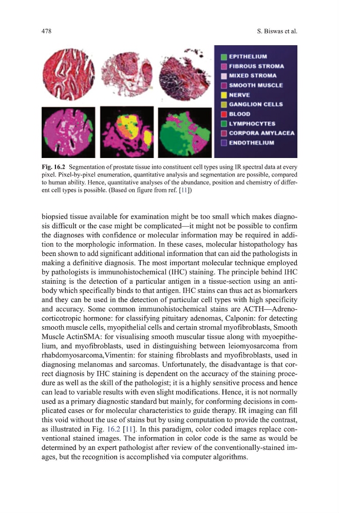
used as a primary diagnostic standard but mainly, for conforming decisions in complicated cases or for molecular characteristics to guide therapy. IR imaging can fill
this void without the use of stains but by using computation to provide the contrast,
as illustrated in Fig. 16.2 [11]. In this paradigm, color coded images replace conventional stained images. the information in color code is the same as would be
determined by an expert pathologist after review of the conventionally-stained images, but the recognition is accomplished via computer algorithms.
Fig. 16.2 Segmentation of prostate tissue into constituent cell types using IR spectral data at every
pixel. Pixel-by-pixel enumeration, quantitative analysis and segmentation are possible, compared
to human ability. hence, quantitative analyses of the abundance, position and chemistry of different cell types is possible. (Based on figure from ref. [11])
S. Biswas et al.
biopsied tissue available for examination might be too small which makes diagnosis difficult or the case might be complicated—it might not be possible to confirm
the diagnoses with confidence or molecular information may be required in addition to the morphologic information. In these cases, molecular histopathology has
been shown to add significant additional information that can aid the pathologists in
making a definitive diagnosis. the most important molecular technique employed
by pathologists is immunohistochemical (IhC) staining. the principle behind IhC
staining is the detection of a particular antigen in a tissue-section using an antibody which specifically binds to that antigen. IhC stains can thus act as biomarkers
and they can be used in the detection of particular cell types with high specificity
and accuracy. Some common immunohistochemical stains are ACth—Adrenocorticotropic hormone: for classifying pituitary adenomas, Calponin: for detecting
smooth muscle cells, myopithelial cells and certain stromal myofibroblasts, Smooth
muscle ActinSmA: for visualising smooth muscular tissue along with myoepithelium, and myofibroblasts, used in distinguishing between leiomyosarcoma from
rhabdomyosarcoma,vimentin: for staining fibroblasts and myofibroblasts, used in
diagnosing melanomas and sarcomas. unfortunately, the disadvantage is that correct diagnosis by IhC staining is dependent on the accuracy of the staining procedure as well as the skill of the pathologist; it is a highly sensitive process and hence
can lead to variable results with even slight modifications. hence, it is not normally
used as a primary diagnostic standard but mainly, for conforming decisions in complicated cases or for molecular characteristics to guide therapy. IR imaging can fill
this void without the use of stains but by using computation to provide the contrast,
as illustrated in Fig. 16.2 [11]. In this paradigm, color coded images replace conventional stained images. the information in color code is the same as would be
determined by an expert pathologist after review of the conventionally-stained images, but the recognition is accomplished via computer algorithms.
Fig. 16.2 Segmentation of prostate tissue into constituent cell types using IR spectral data at every
pixel. Pixel-by-pixel enumeration, quantitative analysis and segmentation are possible, compared
to human ability. hence, quantitative analyses of the abundance, position and chemistry of different cell types is possible. (Based on figure from ref. [11])